Copyright
©The Author(s) 2024.
World J Clin Cases. Jul 6, 2024; 12(19): 4003-4009
Published online Jul 6, 2024. doi: 10.12998/wjcc.v12.i19.4003
Published online Jul 6, 2024. doi: 10.12998/wjcc.v12.i19.4003
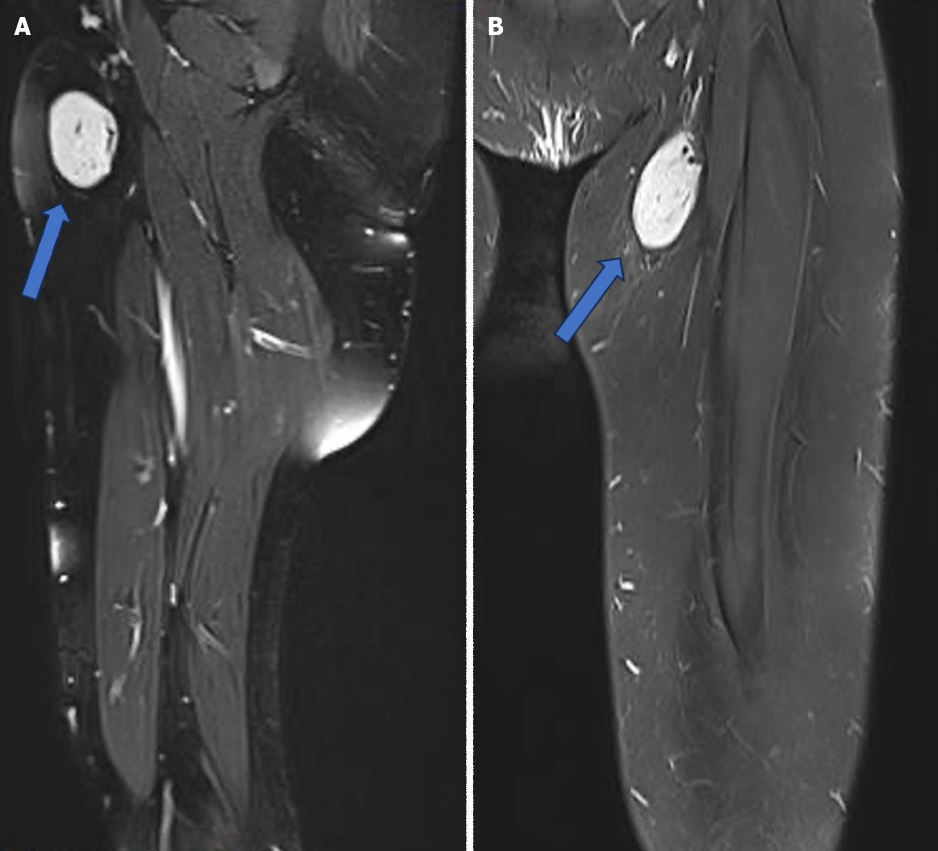
Figure 1 Magnetic resonance imaging showed a well-circumscribed and demarcated cystic lesion projected in the left inguinal region with eccentrically positioned signal void vascular, likely lymphoid in nature (arrow).
A: Sagittal view. B: Coronal view.
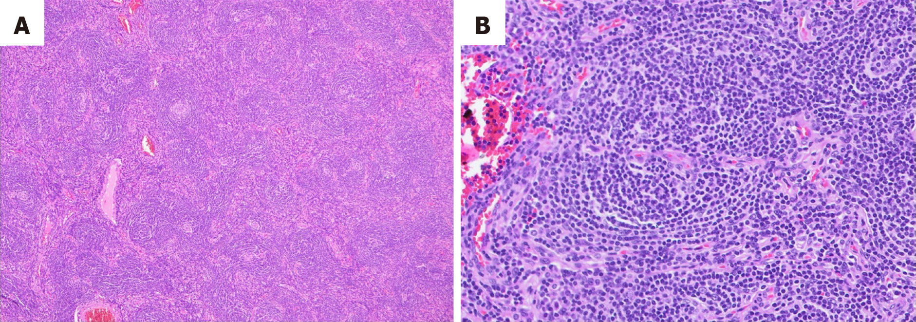
Figure 2 Hematoxylin and eosin staining showing.
A: Regressed follicles with increased endothelial venules; B: Higher magnification of one of the regressed follicles showing a prominent mantle zone (onion skin appearance) and penetrated by vessel.
- Citation: AlSheikh S, Altoijry A, Al-Mubarak H, Alsallum OD, Alakeel F, Alanezi T. A rare presentation of unicentric Castleman's disease in the thigh: A case report and review of literature. World J Clin Cases 2024; 12(19): 4003-4009
- URL: https://www.wjgnet.com/2307-8960/full/v12/i19/4003.htm
- DOI: https://dx.doi.org/10.12998/wjcc.v12.i19.4003


