Copyright
©The Author(s) 2024.
World J Clin Cases. Jul 6, 2024; 12(19): 3791-3799
Published online Jul 6, 2024. doi: 10.12998/wjcc.v12.i19.3791
Published online Jul 6, 2024. doi: 10.12998/wjcc.v12.i19.3791
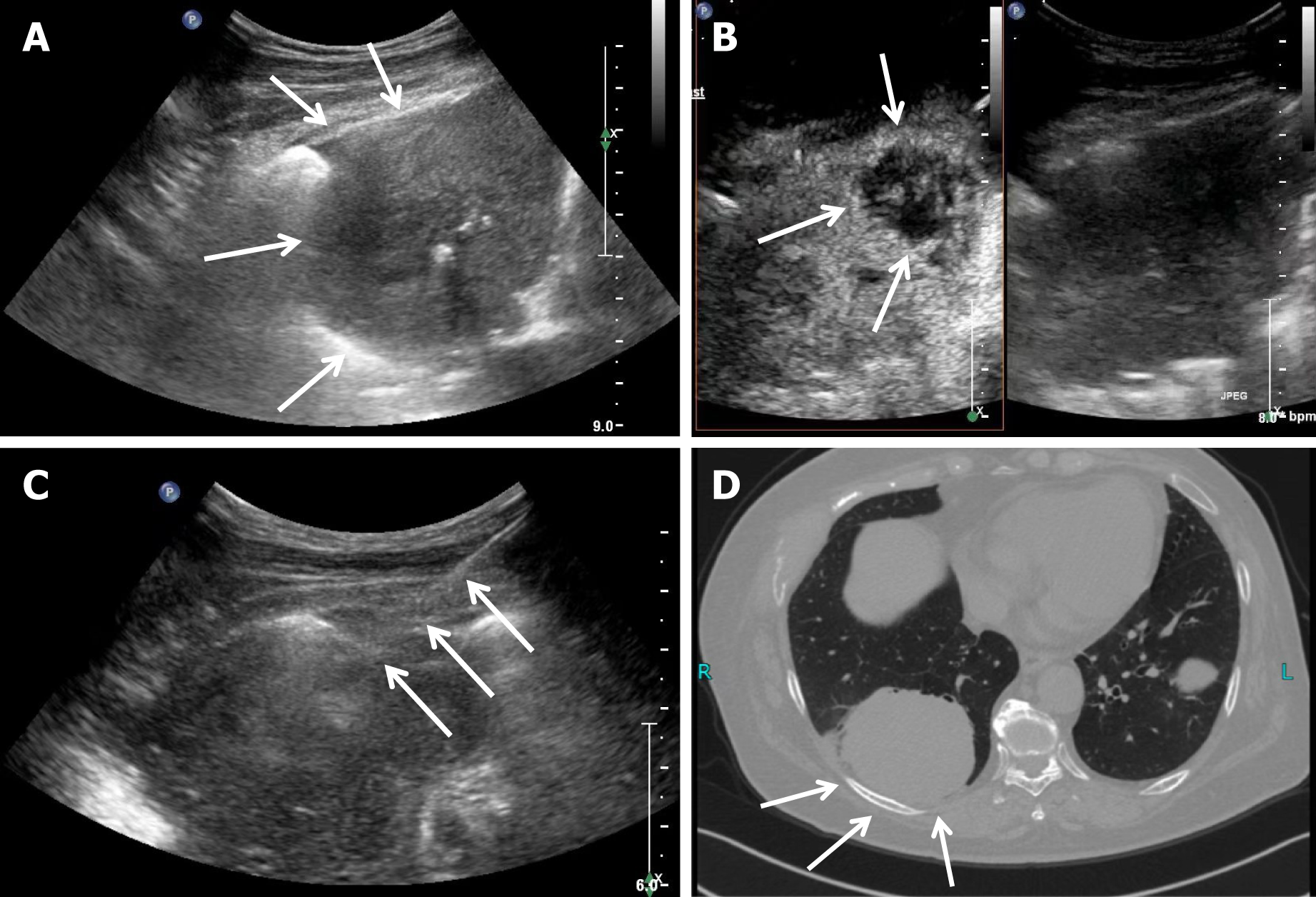
Figure 1 Contrast-enhanced ultrasound examination imaging observation.
A: Large peripheral focal lesion in the right lung (indicated by arrows); B: Contrast-enhanced ultrasound (CEUS) reveals a nonenhanced necrotic area within the peripheral focus (indicated by arrows); C: Puncture needle angle demonstrating avoidance of the nonenhanced necrotic area, as shown by CEUS, and targeting of the region with enhanced activity (indicated by arrows); D: Focal lesions in the posterior basal segment of the right lower lung on computed tomography (indicated by arrows).
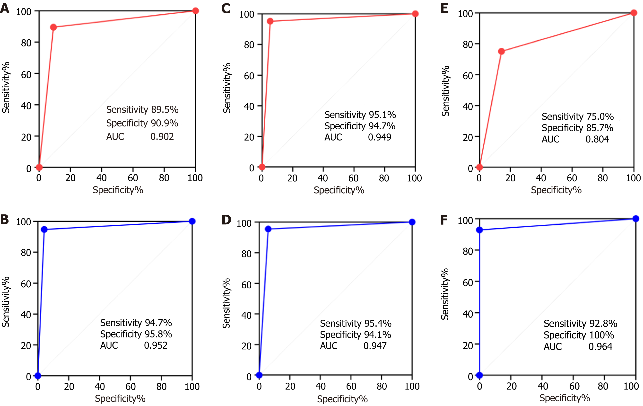
Figure 2 Receiver operating characteristic analysis of the diagnostic efficacy of puncture biopsy for lung carcinomas in different diameter angiography groups and the control group.
A and B: Receiver operating characteristic (ROC), sensitivity, and specificity of the diagnostic efficacy of puncture biopsy in the control and contrast groups; C and D: ROC, sensitivity, and specificity of the diagnostic efficacy of puncture biopsy in the control and contrast groups for lesions 1-5 cm in diameter; E and F: ROC, sensitivity, and specificity of the diagnostic efficacy of puncture biopsy in the control and contrast groups for lesions with diameter ≥ 5 cm. AUC: Area under the curve.
- Citation: Jiang X, Chen J, Gu FF, Li ZR, Song YS, Long JJ, Zhang SZ, Xu TT, Tang YJ, Gu JY, Fang XM. Evaluating the efficacy of percutaneous puncture biopsy guided by contrast-enhanced ultrasound for peripheral pulmonary lesions. World J Clin Cases 2024; 12(19): 3791-3799
- URL: https://www.wjgnet.com/2307-8960/full/v12/i19/3791.htm
- DOI: https://dx.doi.org/10.12998/wjcc.v12.i19.3791


