Copyright
©The Author(s) 2024.
World J Clin Cases. Jun 26, 2024; 12(18): 3609-3614
Published online Jun 26, 2024. doi: 10.12998/wjcc.v12.i18.3609
Published online Jun 26, 2024. doi: 10.12998/wjcc.v12.i18.3609
Figure 1 Colored ultrasound of the thyroid gland and cervical lymph nodes.
A-C: The left lobe of the thyroid gland is enlarged, the right lobe is not large, and the echogenicity of glandular tissue is uneven. A mixed echogenic mass of about 38 mm × 30 mm was found in the left lobe of the thyroid, with a clear boundary and regular shape. Several strong echogenic spots of different sizes were observed inside, accompanied by acoustic shadow behind.
Figure 2 Parathyroid imaging (MIBI).
A-C: A concentrated shadow of radioactivity in the left lobe of the thyroid.
Figure 3 Fine needle aspiration cytological smear of thyroid nodules.
A-C: Patches of thyroid follicular epithelium with crowded cells were detected. Some cells showed intranuclear pseudoinclusions, and nuclear furrows were rare. The pathological case was considered papillary thyroid carcinoma.
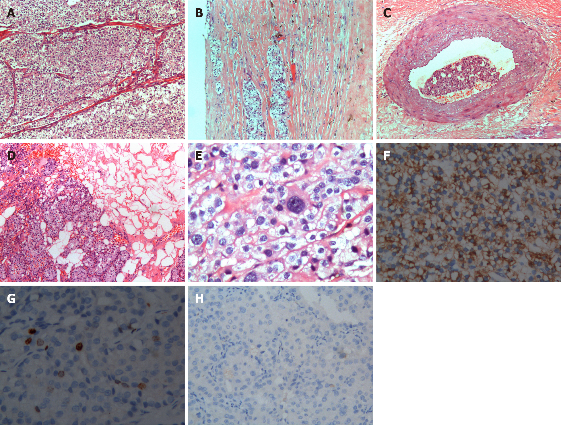
Figure 4 Pathological findings.
A: Fiber separation (10×); B: Capsular invasion (10×); C: Endovascular invasion (10×); D: Fatty infiltration (10×). E: Heterocyst (40×); F: Parathyroid hormone staining (40×); G: Ki67 staining (40×); H: Calcitonin staining (40×).
- Citation: Gui SY, Zhang CN, Ling L, Tang RX, Yang J. Parathyroid carcinoma located in the thyroid gland: A case report. World J Clin Cases 2024; 12(18): 3609-3614
- URL: https://www.wjgnet.com/2307-8960/full/v12/i18/3609.htm
- DOI: https://dx.doi.org/10.12998/wjcc.v12.i18.3609


