Copyright
©The Author(s) 2024.
World J Clin Cases. Jun 16, 2024; 12(17): 3271-3276
Published online Jun 16, 2024. doi: 10.12998/wjcc.v12.i17.3271
Published online Jun 16, 2024. doi: 10.12998/wjcc.v12.i17.3271
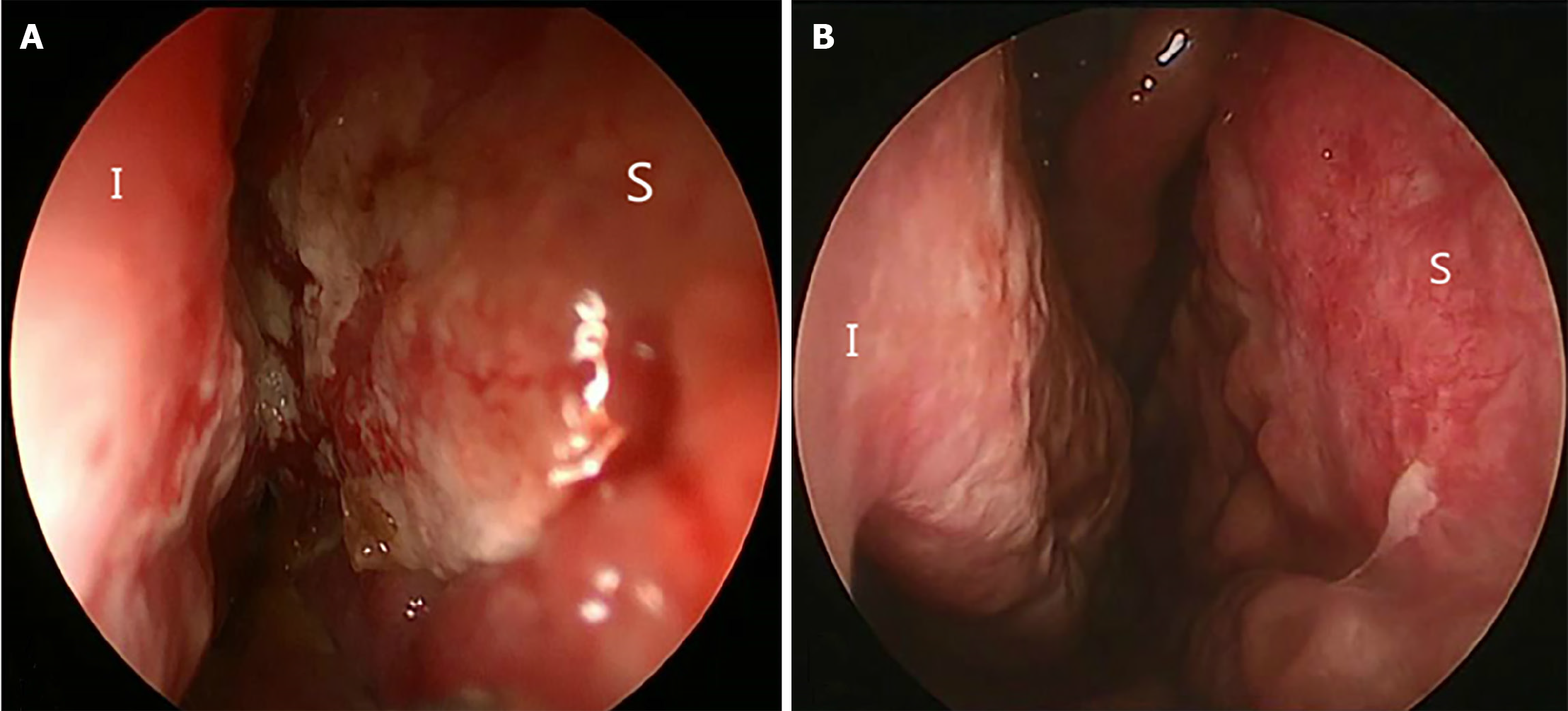
Figure 1 Endoscopy images.
A: Pre-treatment endoscopic finding. The nasal mucosa was thickened, inflamed and crusting; B: Post-treatment endoscopic finding. The nasal mucosa gradually became smooth.
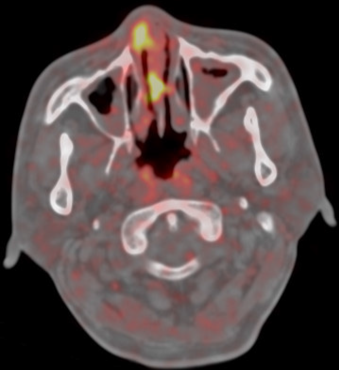
Figure 2 Pathological hypercaptation of the right nasal cavity and septum in 18F-fluorodeoxyglucose positron emission tomography/computed tomography.
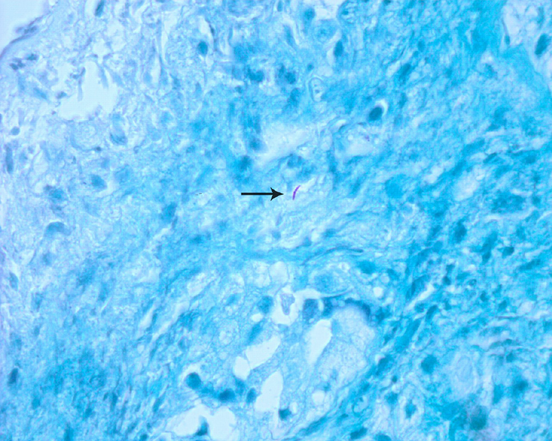
Figure 3 Ziehl-Neelsen staining × 1000, demonstrating acid fast bacilli.
- Citation: Liu YC, Zhou ML, Cheng KJ, Zhou SH, Wen X. Treatment of primary nasal tuberculosis with anti-tumor necrosis factor immunotherapy: A case report. World J Clin Cases 2024; 12(17): 3271-3276
- URL: https://www.wjgnet.com/2307-8960/full/v12/i17/3271.htm
- DOI: https://dx.doi.org/10.12998/wjcc.v12.i17.3271


