Copyright
©The Author(s) 2024.
World J Clin Cases. Jun 16, 2024; 12(17): 3259-3264
Published online Jun 16, 2024. doi: 10.12998/wjcc.v12.i17.3259
Published online Jun 16, 2024. doi: 10.12998/wjcc.v12.i17.3259
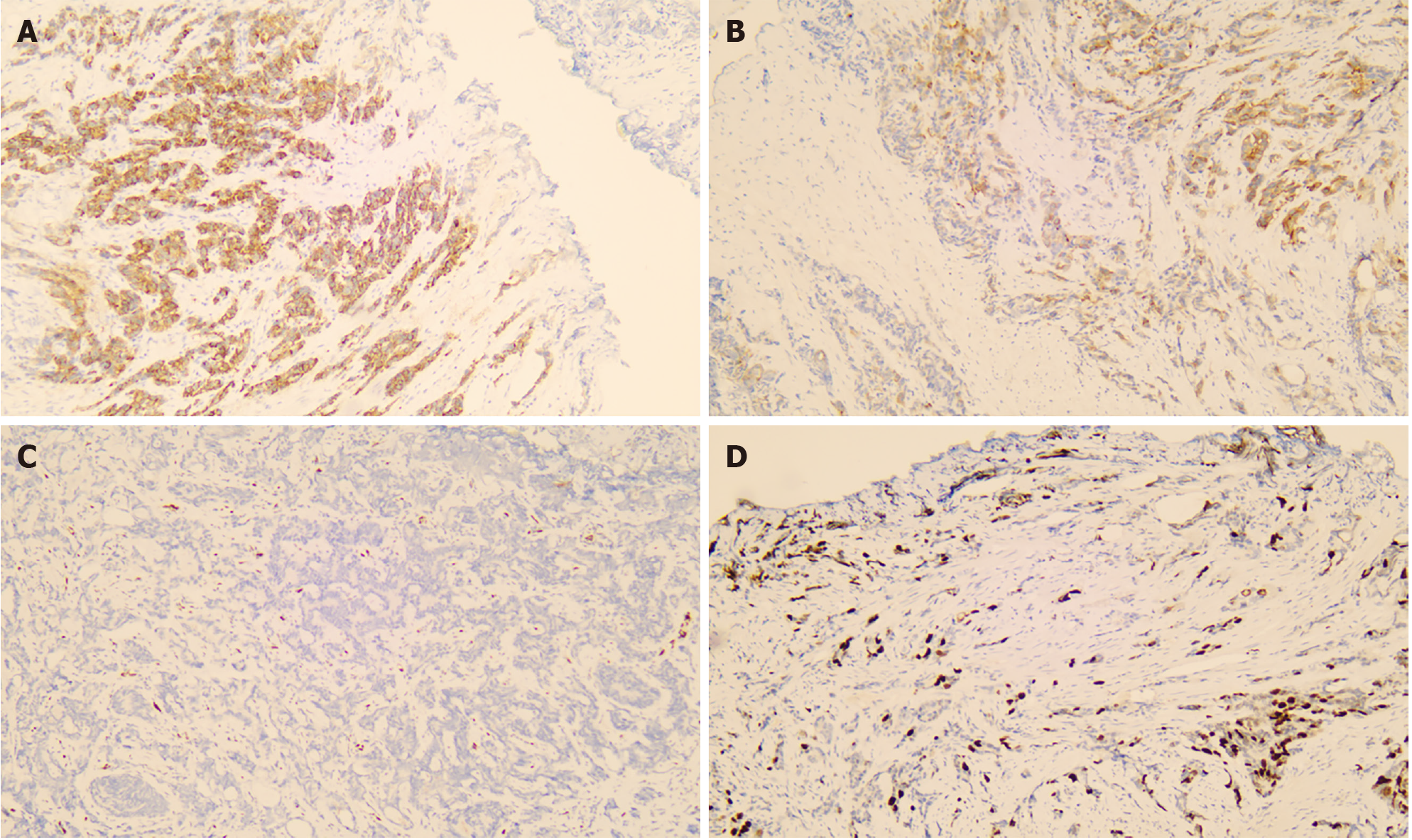
Figure 1 Histopathological features of the surgically removed prostate.
A: P504S-positive, immunohistochemical staining; B: Prostate-specific-antigen-positive, immunohistochemical staining; C: Early growth response gene-positive, immunohistochemical staining; D: Ki67-positive, immunohistochemical staining (all images magnified 40 ×).
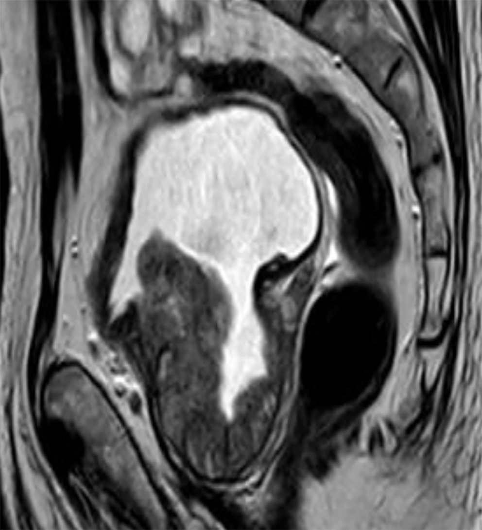
Figure 2 Schematic diagram of magnetic resonance imaging.
The prostatic protrusion was evident on T2 weighted imagin.
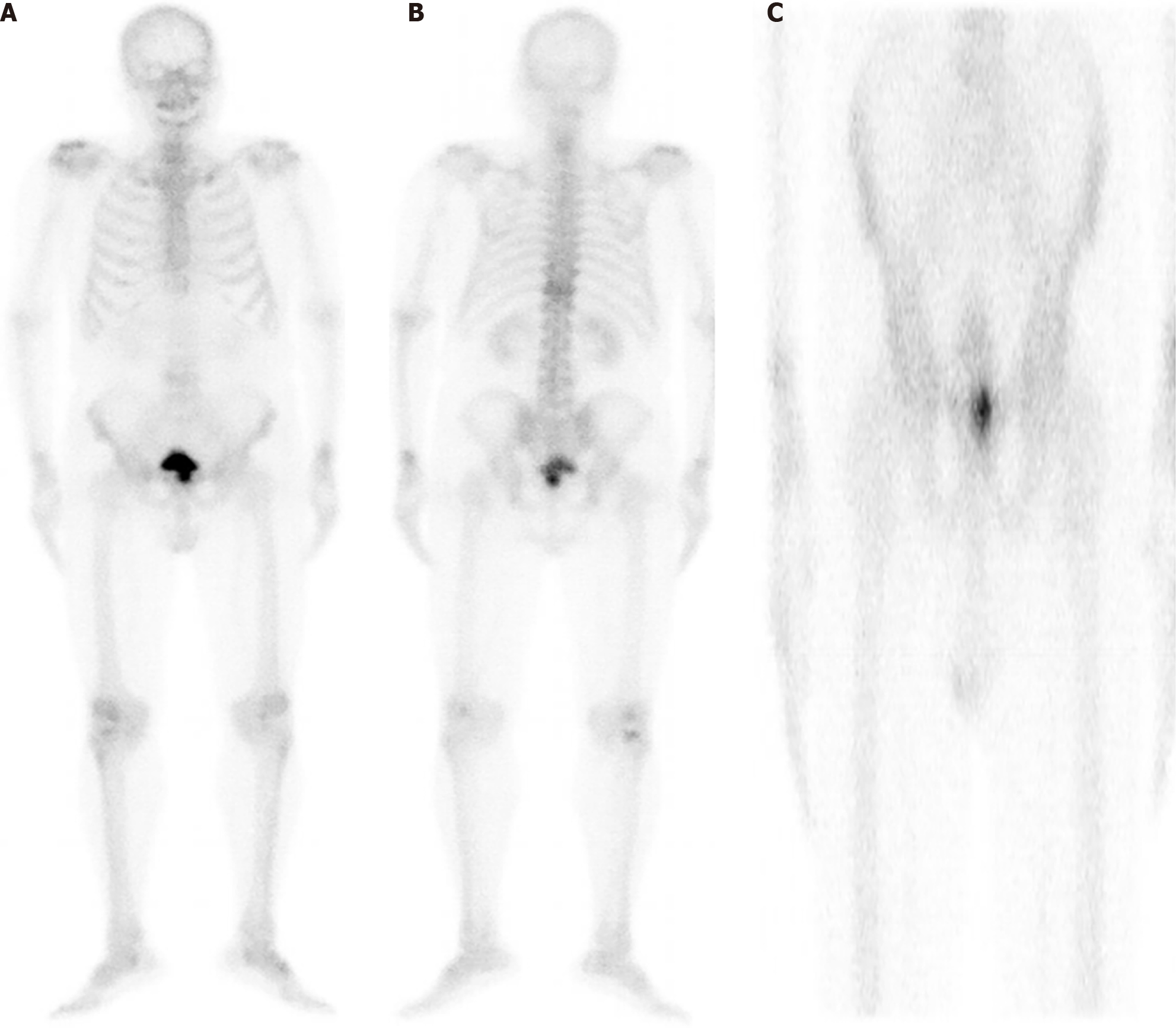
Figure 3 Imaging was performed after intravenous injection of 99mTc-MDP.
A: Whole body bone anterior position; B: The whole body is posterior; C: Local fault.
- Citation: Huang DH, Hu YX, Guo S, Yang WJ. Prostate cancer with elevated free prostate-specific antigen density: A case report. World J Clin Cases 2024; 12(17): 3259-3264
- URL: https://www.wjgnet.com/2307-8960/full/v12/i17/3259.htm
- DOI: https://dx.doi.org/10.12998/wjcc.v12.i17.3259


