Copyright
©The Author(s) 2024.
World J Clin Cases. Jun 16, 2024; 12(17): 3206-3213
Published online Jun 16, 2024. doi: 10.12998/wjcc.v12.i17.3206
Published online Jun 16, 2024. doi: 10.12998/wjcc.v12.i17.3206
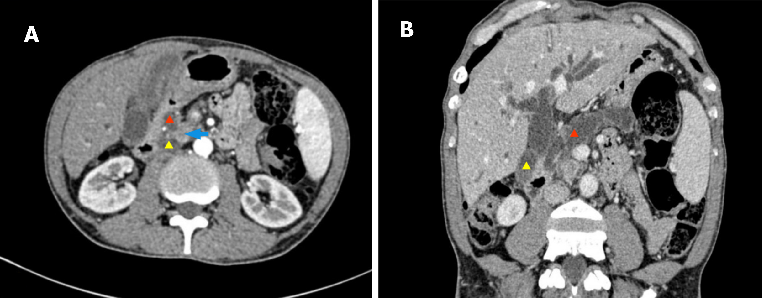
Figure 1 Enhanced abdominal computed tomography scan of case 1.
A nodular shadow (blue arrow) is visible in the pancreatic head region, with significant dilation of the intrahepatic and extrahepatic bile ducts. The local pancreatic duct is interrupted at the lesion site, and the distal pancreatic duct is significantly dilated. The pancreatic parenchyma shows atrophy. Red triangle: Main pancreatic duct; Yellow triangle: Common bile duct. A: Axial phase; B: Reconstruction of the biliary and pancreatic duct system.
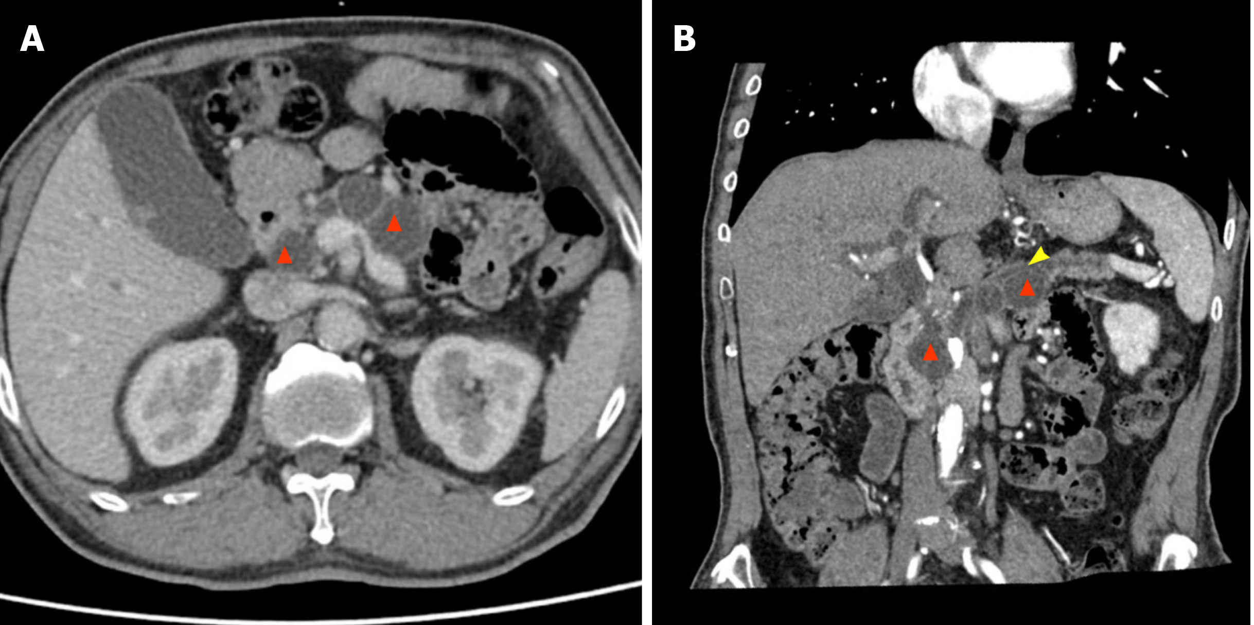
Figure 2 Enhanced abdominal computed tomography scan of case 2.
Multiple cystic low-density shadows are visible in the pancreatic head and body, with pancreatic parenchymal atrophy and dilation of the main pancreatic duct. Red triangle: Multiple cystic masses in the pancreatic head and body; Yellow arrow: Dilation of the main pancreatic duct. A: Axial phase; B: Reconstruction of the biliary and pancreatic duct system.
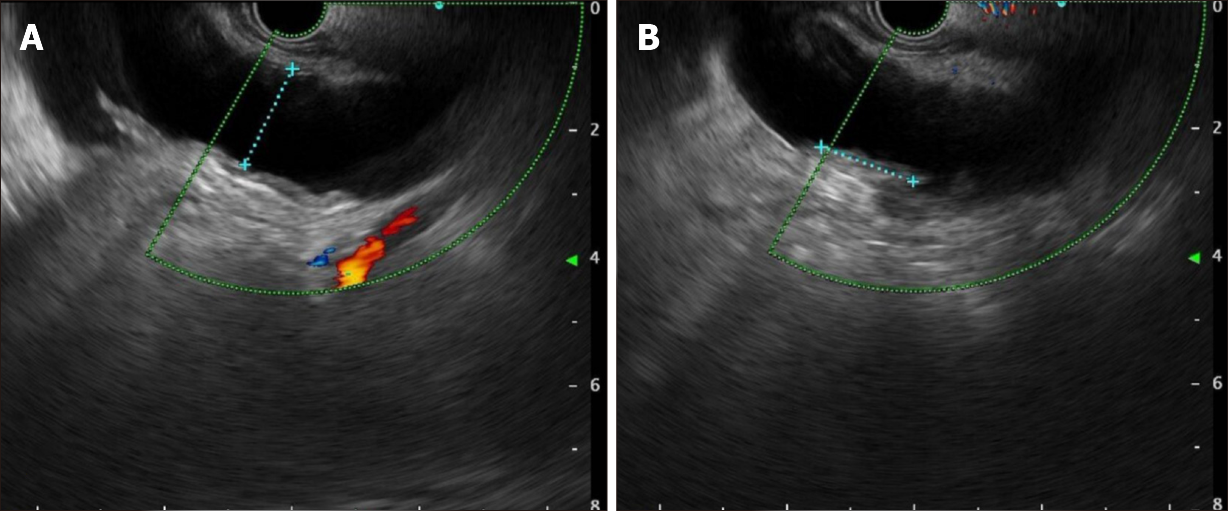
Figure 3 Endoscopic ultrasound images of case 1.
A: Significant dilation of the main pancreatic duct, approximately 1.7 cm; B: Isoechoic nodules in the ductal wall.
Figure 4 Surgical schematic diagram for case 1.
A: Before resection; B: After resection and reconstruction. SpA: Splenic artery; SpV: Splenic vein; PV: Portal vein; SMV: Superior mesenteric vein.
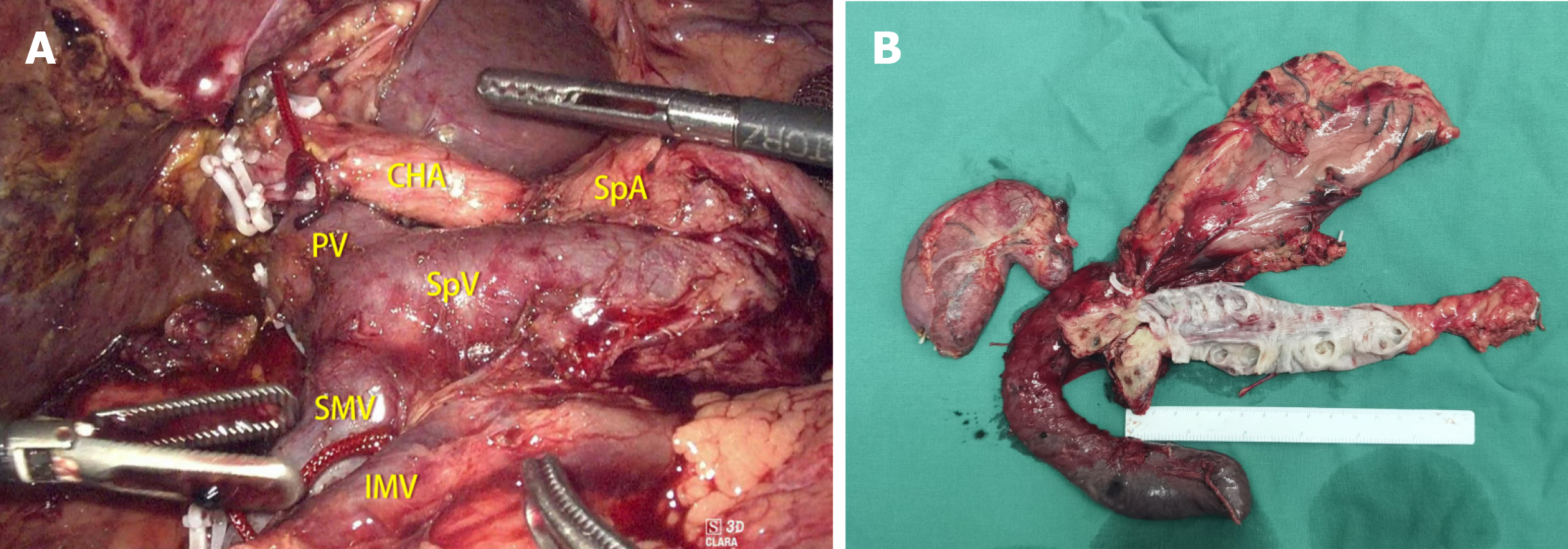
Figure 5 Tumor information for case 1.
A: Intraoperative view after resection of the tumor in case 1; B: Gross specimen from case 1. CHA: Common hepatic artery; SpA: Splenic artery; SpV: Splenic vein; PV: Portal vein; SMV: Superior mesenteric vein; IMV: Inferior mesenteric vein.
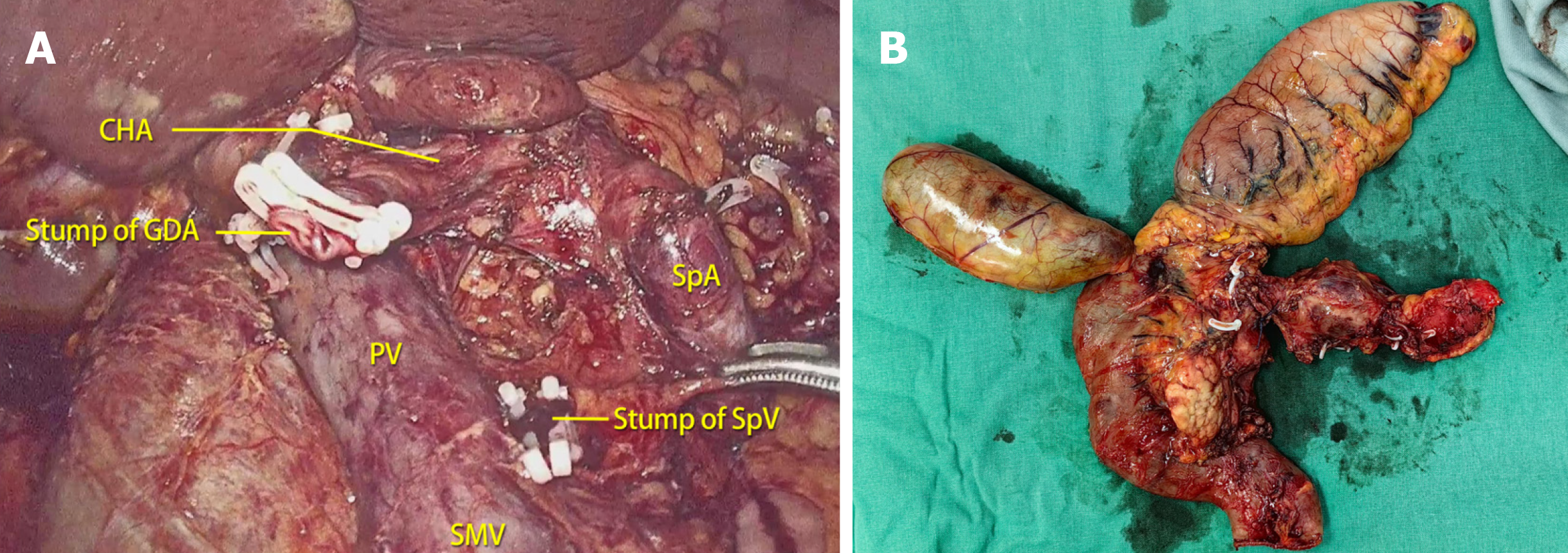
Figure 6 Tumor information for case 2.
A: Intraoperative view after resection of the tumor in case 2; B: Gross specimen from case 2. CHA: Common hepatic artery; GDA: Gastroduodenal artery; SpA: Splenic artery; SpV: Splenic vein; PV: Portal vein; SMV: Superior mesenteric vein.
- Citation: Sun MQ, Kang XM, He XD, Han XL. Laparoscopic spleen-preserving total pancreatectomy for the treatment of low-grade malignant pancreatic tumors: Two case reports and review of literature. World J Clin Cases 2024; 12(17): 3206-3213
- URL: https://www.wjgnet.com/2307-8960/full/v12/i17/3206.htm
- DOI: https://dx.doi.org/10.12998/wjcc.v12.i17.3206


