Copyright
©The Author(s) 2024.
World J Clin Cases. Jun 16, 2024; 12(17): 3138-3143
Published online Jun 16, 2024. doi: 10.12998/wjcc.v12.i17.3138
Published online Jun 16, 2024. doi: 10.12998/wjcc.v12.i17.3138
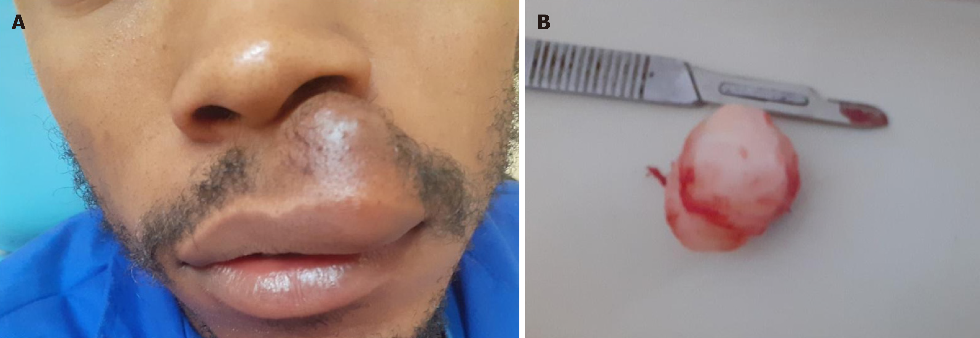
Figure 1 View of the patient and surgical specimen.
A: Profile view of the patient on presentation; B: Surgical specimen.
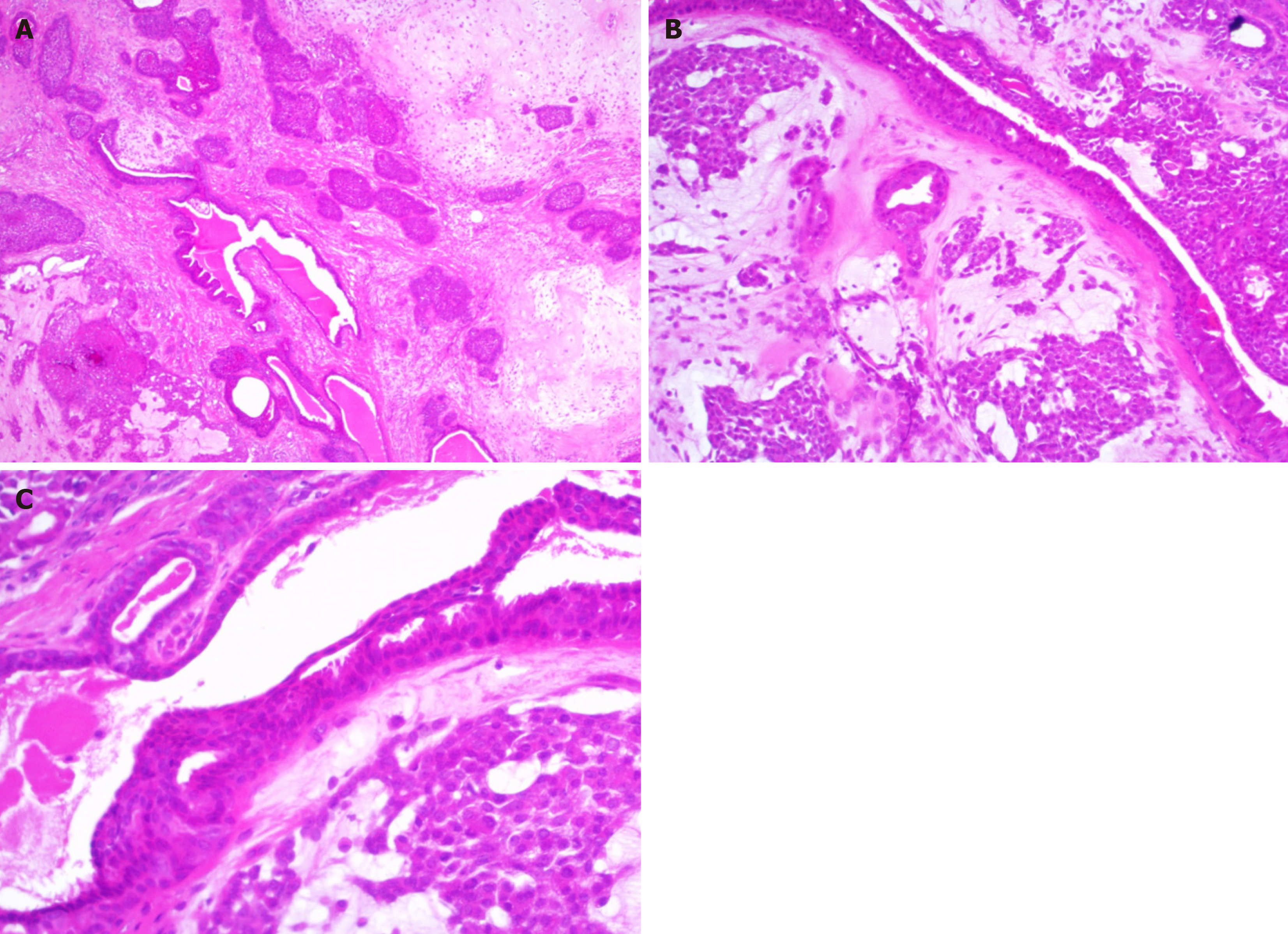
Figure 2 Epithelial nests and tubules associated with sheets and clusters of plasmacytoid myoepithelial cells embedded in a chondromyxoid matrix.
A: Haematoxylin and eosin (H&E) staining, × 40 magnification; B: H&E staining, × 100 magnification; C: H &E staining, × 200 magnification.
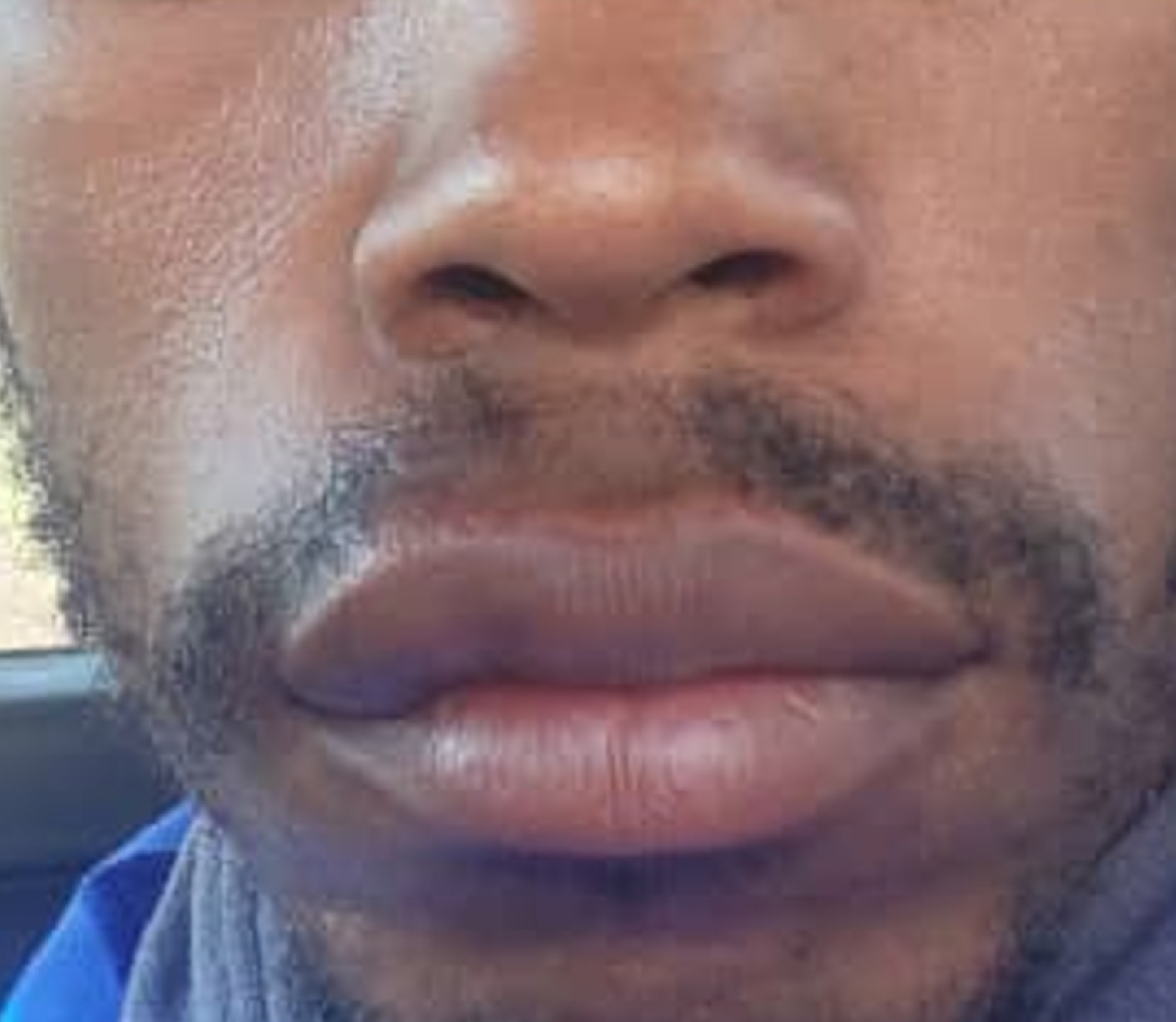
Figure 3 View of the patient 6 wk post-surgery.
- Citation: Chidzonga MM, Mahomva L, Zambuko B, Muungani W. Pleomorphic adenoma (mixed tumor) of the upper lip: A case report. World J Clin Cases 2024; 12(17): 3138-3143
- URL: https://www.wjgnet.com/2307-8960/full/v12/i17/3138.htm
- DOI: https://dx.doi.org/10.12998/wjcc.v12.i17.3138


