Copyright
©The Author(s) 2024.
World J Clin Cases. Apr 26, 2024; 12(12): 2079-2085
Published online Apr 26, 2024. doi: 10.12998/wjcc.v12.i12.2079
Published online Apr 26, 2024. doi: 10.12998/wjcc.v12.i12.2079
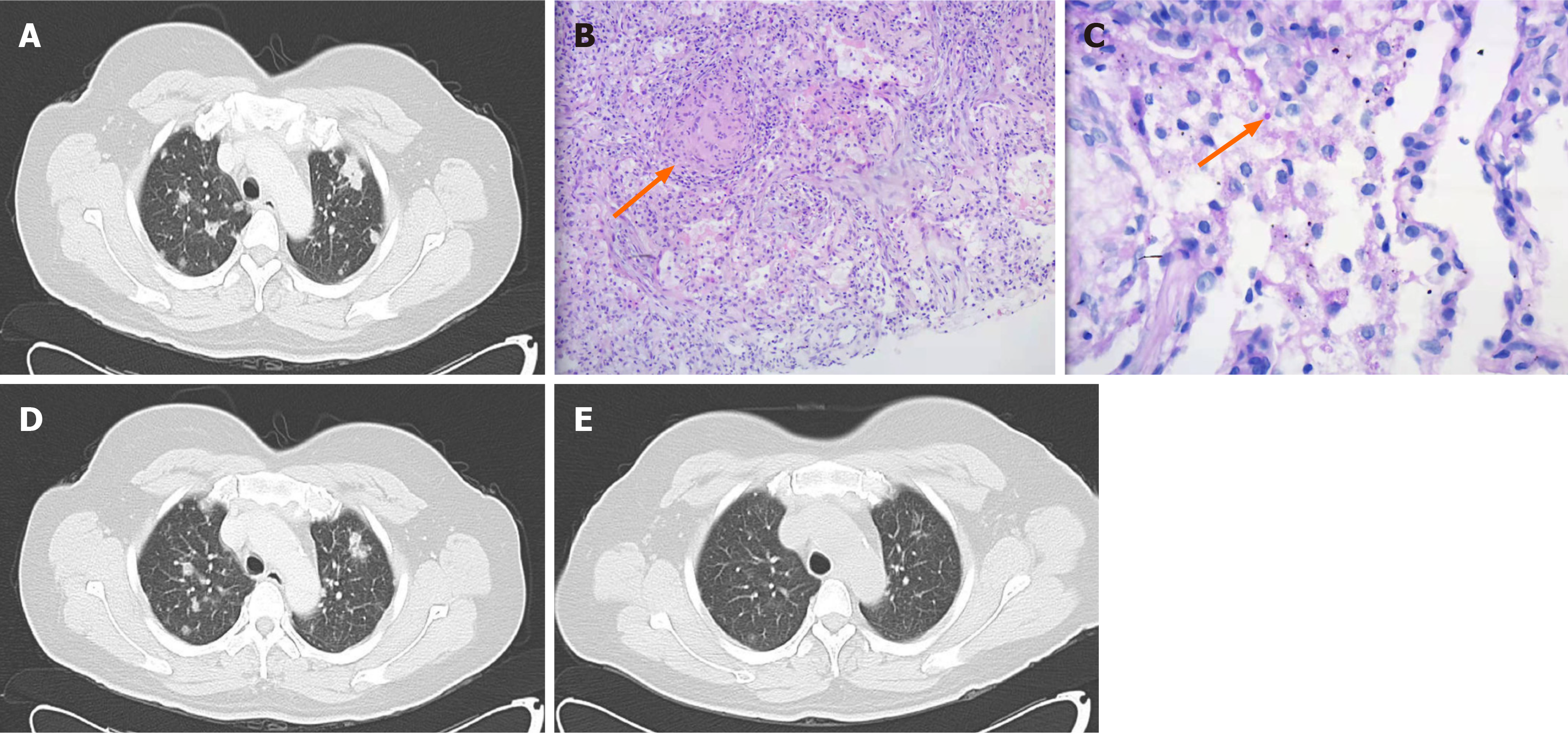
Figure 1 Changes of chest computed tomography findings and histopathology results.
A: Scan at admission, revealing multiple nodules of unequal size with uneven internal density and multiple small burrs at the edges of the lesions; B: Pathology of pulmonary nodules, suggesting inflammation and granuloma formation; C: Pathology of pulmonary nodules, revealing a yeast-like corpuscle in the alveolar cavity; D: Scan after 2 wk of antibiotic treatment, showing the nodules were significantly reduced; E: Scan after 1 month of antibiotic treatment, showing the pulmonary lesions had obviously disappeared.
- Citation: Huang HY, Bu KP, Liu JW, Wei J. Overlapping infections of Mycobacterium canariasense and Nocardia farcinica in an immunocompetent patient: A case report. World J Clin Cases 2024; 12(12): 2079-2085
- URL: https://www.wjgnet.com/2307-8960/full/v12/i12/2079.htm
- DOI: https://dx.doi.org/10.12998/wjcc.v12.i12.2079


