Copyright
©The Author(s) 2024.
World J Clin Cases. Apr 16, 2024; 12(11): 1954-1959
Published online Apr 16, 2024. doi: 10.12998/wjcc.v12.i11.1954
Published online Apr 16, 2024. doi: 10.12998/wjcc.v12.i11.1954
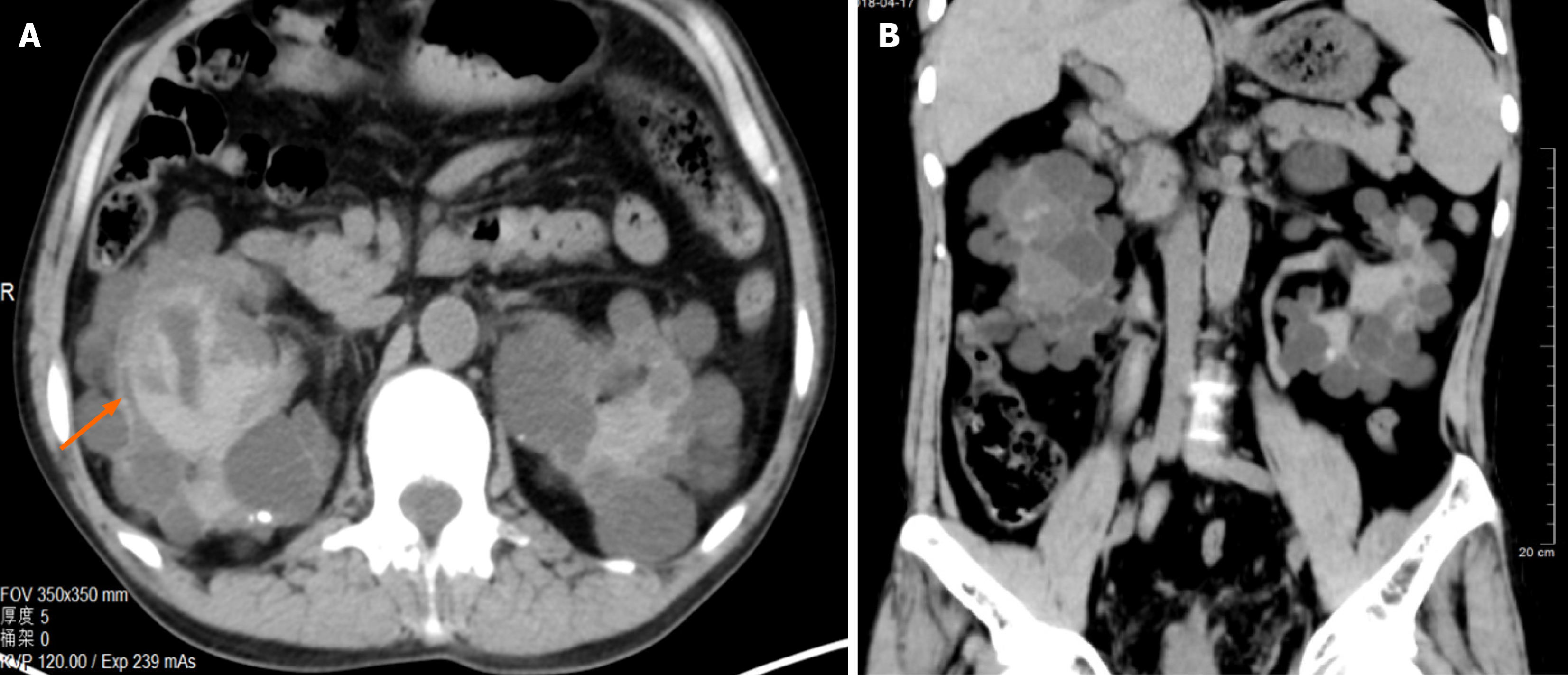
Figure 1 51-year-old male patient with autosomal dominant polycystic kidney disease had gross hematuria for 1 wk.
He regularly conducts hemodialysis in hospital. The abdominal no-contrast computerized tomography scan showed bilateral enlarged kidneys which were full of cysts of uniform size. Acute hemorrhage was found in the cysts (orange arrow).
Figure 2 Bilateral renal arteriography.
A: Bilateral renal arteriography showed bilateral enlarged kidneys and slender branches of renal arteries with contrast-medium leaking indicating acute hemorrhage (orange arrows); B: Bilateral renal arteriography showed bilateral enlarged kidneys with microcoils positioned at the main renal arterials and contrast-medium leaking indicating acute hemorrhage (orange arrows).
Figure 3 Autosomal dominant polycystic kidney disease patients.
Bilateral renoarteriography showed that microcoils positioned at the renal arterial branches and no contrast-medium leaking indicating the success of hemostasis (orange arrows). A: Conducted transcatheter renal artery embolization with microspheres of polyvinyl alcohol (diameter, 350 μm-560 μm) in branches of renal arteries and microcoils; B: Conducted transcatheter renal artery embolization with microspheres of polyvinyl alcohol (diameter, 350 μm-560 μm) in branches of renal arteries and microcoils in main renal arteries.
- Citation: Sui WF, Duan YX, Li JY, Shao WB, Fu JH. Safety and efficacy of transcatheter arterial embolization in autosomal dominant polycystic kidney patients with gross hematuria: Six case reports. World J Clin Cases 2024; 12(11): 1954-1959
- URL: https://www.wjgnet.com/2307-8960/full/v12/i11/1954.htm
- DOI: https://dx.doi.org/10.12998/wjcc.v12.i11.1954


