Copyright
©The Author(s) 2024.
World J Clin Cases. Apr 6, 2024; 12(10): 1785-1792
Published online Apr 6, 2024. doi: 10.12998/wjcc.v12.i10.1785
Published online Apr 6, 2024. doi: 10.12998/wjcc.v12.i10.1785
Figure 1 Radiographs at first hospitalization at the age of 5.
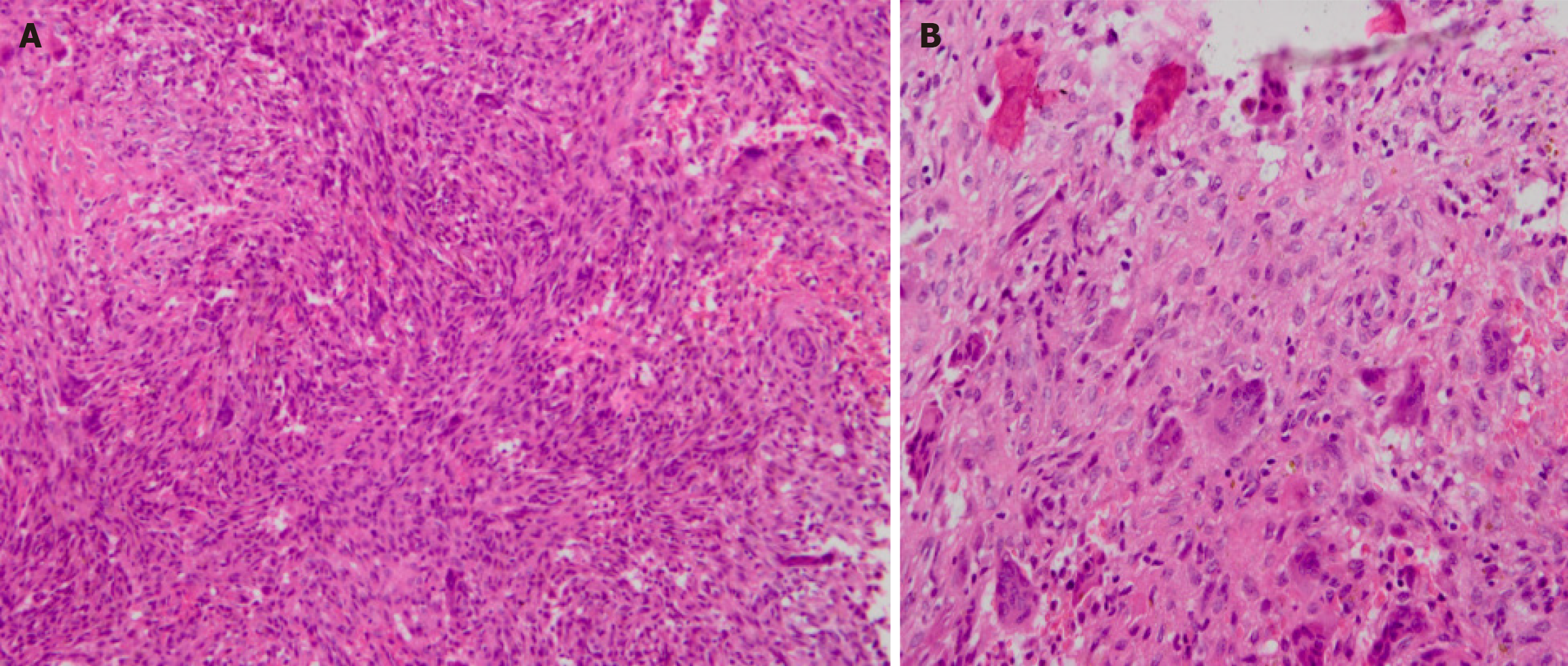
Figure 2 Histological findings.
A: × 200; B: × 400. Histopathological examination showed spindle-shaped fibroblastic and collagenous stromal tissue, characteristic of non-ossifying fibroma, then diagnosis of Jaffe-Campanacci syndrome was made. Conservative therapy was applied with suggestions of observation, reduction of mobility and follow-up.
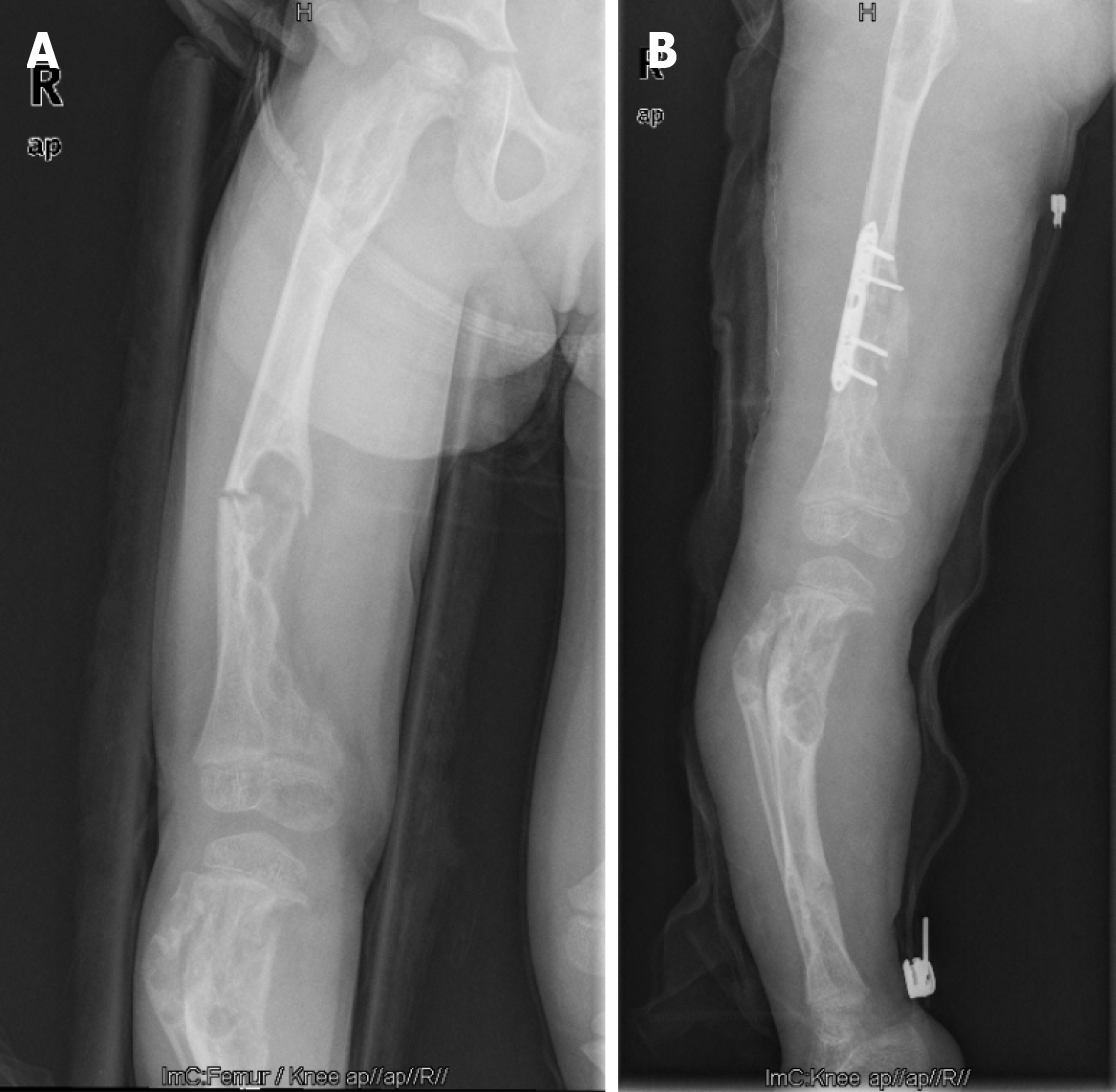
Figure 3 Patient at the age of 7: X-rays before and after surgery.
A: X-rays before surgery; B: X-rays after surgery.
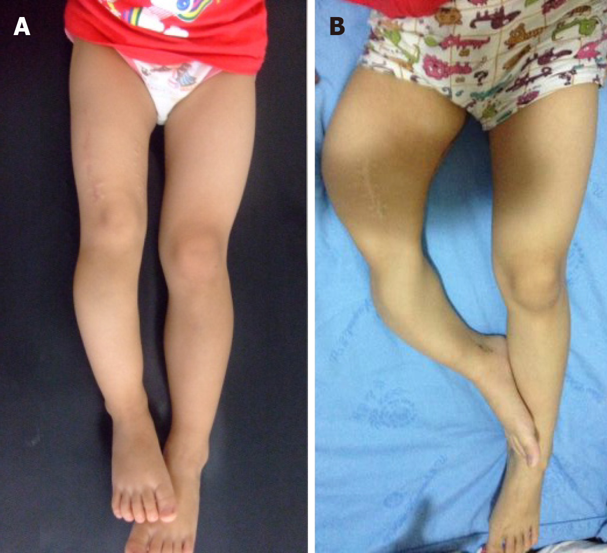
Figure 4 Changes in the appearance of both lower limbs.
A: Patient at age 5, after curettage and biopsy of the distal part of the right femur: The right leg is about 5 cm shorter than the left leg; B: Patient at age 10, before amputation: The right leg is obviously shorter than the left side and shows severe deformity of the thigh and shank.
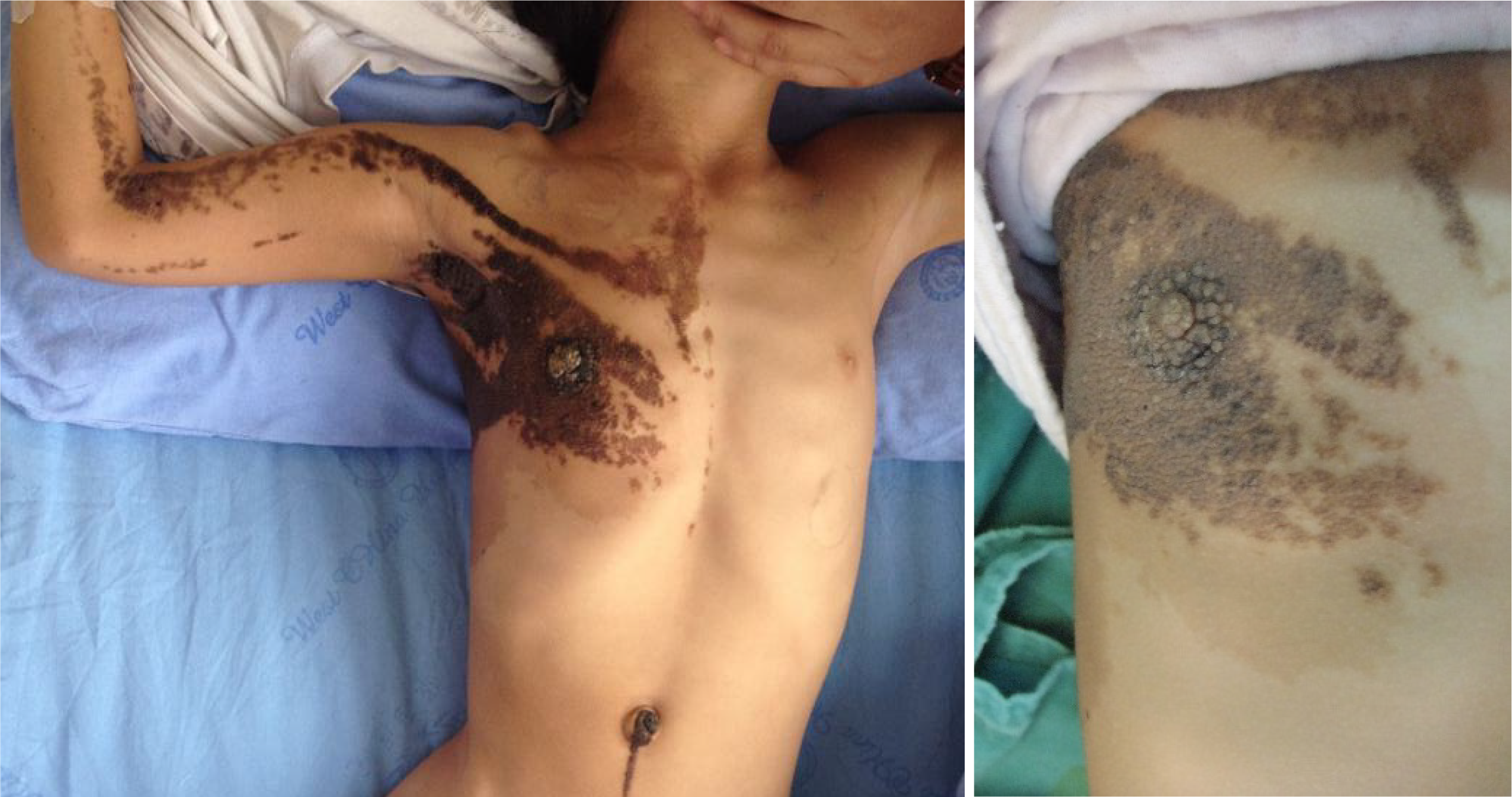
Figure 5 Multiple café-au-lait macules were noted on the child’s body.
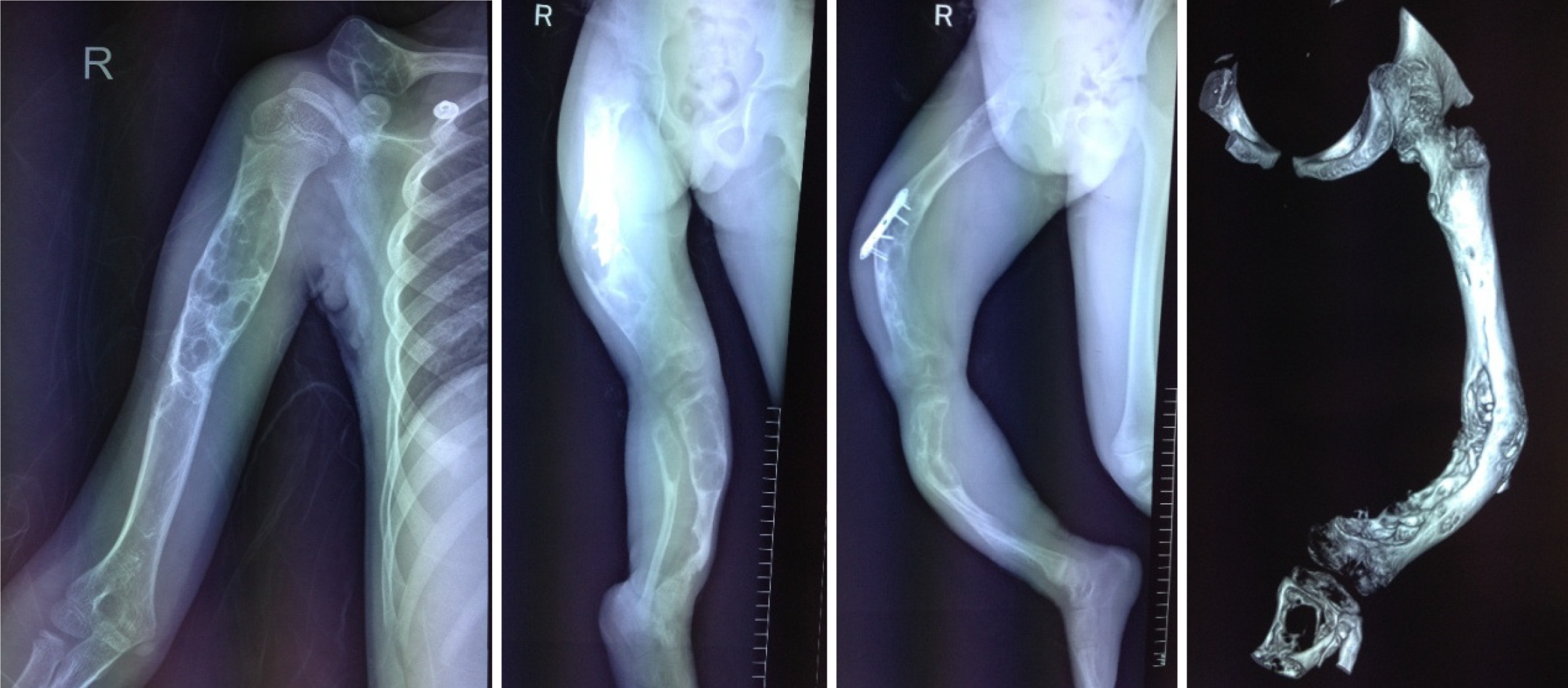
Figure 6 Radiographs of the right humerus, right clavicle, right femur, right tibia, and fibula before amputation.
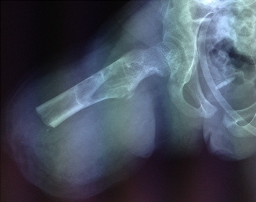
Figure 7 X-ray of the right femur 1 d after amputation.
- Citation: Jiang J, Liu M. Jaffe-Campanacci syndrome resulted in amputation: A case report. World J Clin Cases 2024; 12(10): 1785-1792
- URL: https://www.wjgnet.com/2307-8960/full/v12/i10/1785.htm
- DOI: https://dx.doi.org/10.12998/wjcc.v12.i10.1785


