Copyright
©The Author(s) 2023.
World J Clin Cases. Mar 26, 2023; 11(9): 2021-2028
Published online Mar 26, 2023. doi: 10.12998/wjcc.v11.i9.2021
Published online Mar 26, 2023. doi: 10.12998/wjcc.v11.i9.2021
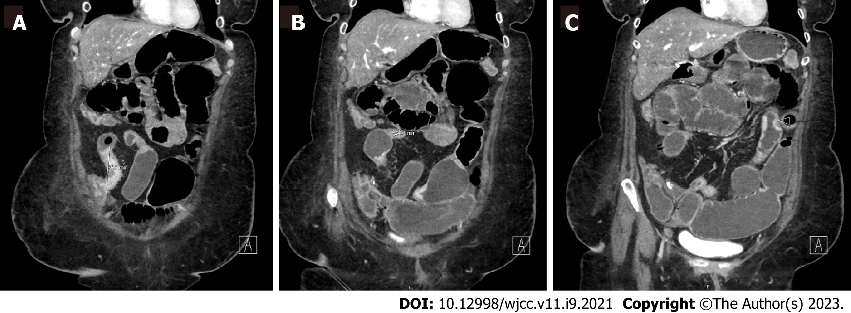
Figure 1 Computed tomography enterography demonstrating multifocal stricturing Crohn’s disease.
A: Persistent areas of mucosal hyperenhancement and segments of stricturing throughout the small bowel. This included an approximately 6.5 cm area of stricturing of the terminal ileum just proximal to the ileocecal valve; B: Additional smaller segments of stricturing of the terminal ileum proximal to this measuring approximately 2.8 cm and 1.8 cm; C: Additional more proximal 4.5 cm segment of stricturing. Possible short segments of stricturing of jejunum in the left hemiabdomen. No fistulas visualized.
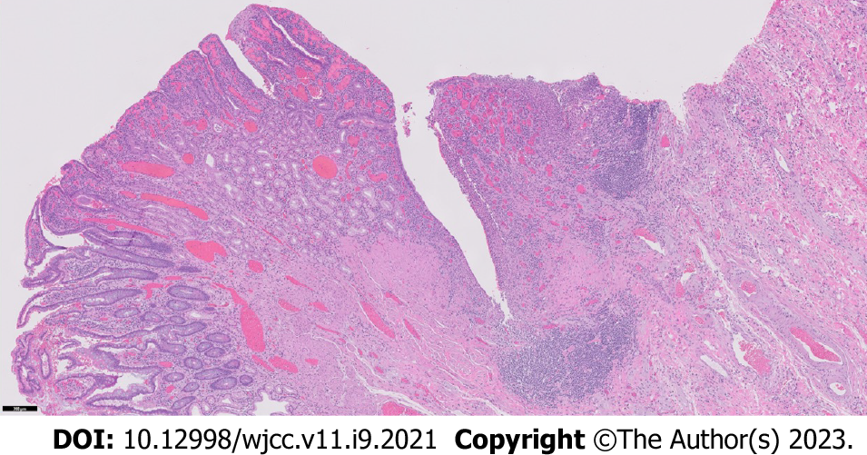
Figure 2 Resected ileum with severe chronic active Crohn’s ileitis.
The background ileum showed severe chronic active ileitis with cryptitis, crypt abscess formation, fissuring ulceration (center), architectural distortion, pyloric metaplasia, and lymphoid aggregates (5 × objective magnification).
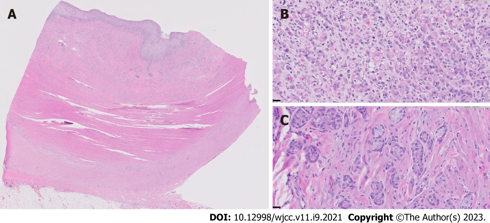
Figure 3 Small bowel adenocarcinoma in resected neoterminal ileum.
A: Cross section of the ileum near the anastomosis, showing complete effacement of the mucosa and infiltration of the underlying ileal wall by the adenocarcinoma (0.5 × objective magnification); B: Representative high-magnification view of the poorly-differentiated adenocarcinoma, comprised of sheets of single malignant cells and cell clusters with signet ring morphology (40 × objective magnification); C: Representative high-magnification view of a well-differentiated region of the adenocarcinoma, characterized by well-defined tubules with small, rigid lumina, mimicking intestinal crypts, as well as tumor cells arranged in well-polarized clusters without distinct lumina (40 × objective magnification).Tumor was noted in 6/35 lymph nodes and showed serosal involvement and lymphovascular and perineural invasion. Immunohistochemistry revealed intact expression of DNA mismatch proteins MLH1, MSH2, MSH6 and PMS2.
- Citation: Karthikeyan S, Shen J, Keyashian K, Gubatan J. Small bowel adenocarcinoma in neoterminal ileum in setting of stricturing Crohn’s disease: A case report and review of literature . World J Clin Cases 2023; 11(9): 2021-2028
- URL: https://www.wjgnet.com/2307-8960/full/v11/i9/2021.htm
- DOI: https://dx.doi.org/10.12998/wjcc.v11.i9.2021


