Copyright
©The Author(s) 2023.
World J Clin Cases. Mar 6, 2023; 11(7): 1615-1626
Published online Mar 6, 2023. doi: 10.12998/wjcc.v11.i7.1615
Published online Mar 6, 2023. doi: 10.12998/wjcc.v11.i7.1615
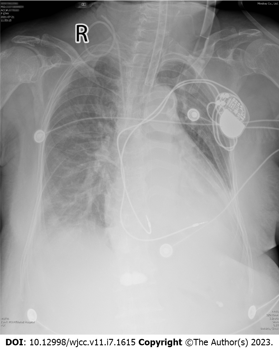
Figure 1 Chest X-ray of the patient on intensive care unit admission.
Chest X-ray performed on intensive care unit admission revealed a mildly enlarged heart and proper positioning of both pacemaker leads.
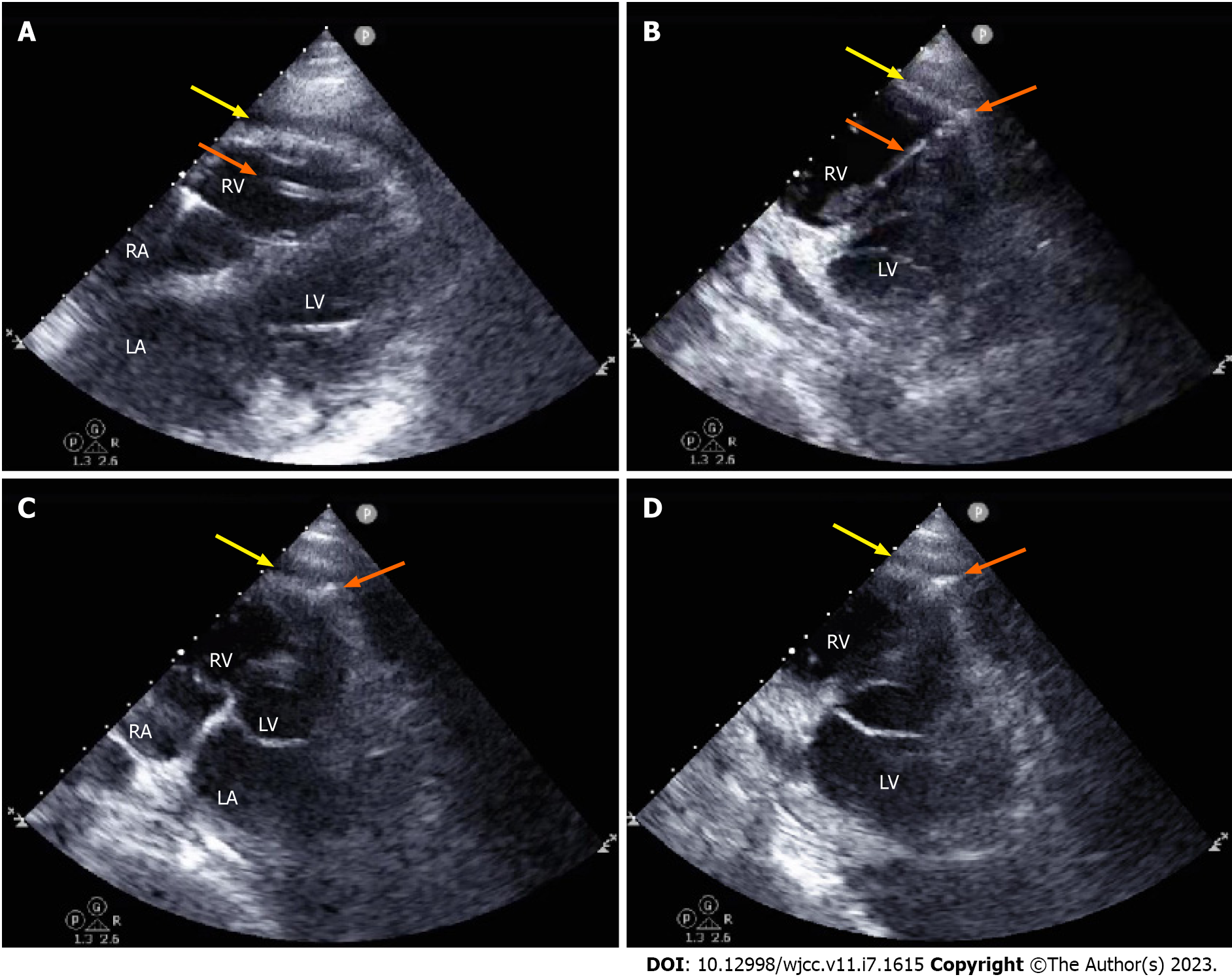
Figure 2 Imaging manifestations on point-of-care ultrasound indicating pacemaker lead perforation of the right ventricle.
A: Parasternal four-chamber view showing pericardial effusion and the outline of a pacemaker lead in the right ventricle (RV) chamber; B: Nonstandard apical RV view showing pericardial effusion and the lead with its tip penetrating the RV wall at the apex, producing a “bow-and-arrow” sign; C and D: Apical four-chamber view (C) and nonstandard apical RV view (D) focusing on the tip and showing the lead penetrating the apical wall and projecting into the free pericardial space. Yellow arrows denote pericardial effusion and orange arrows denote the pacemaker lead; RA: Right atrium; RV: Right ventricle; LA: Left atrium; LV: Left ventricle.
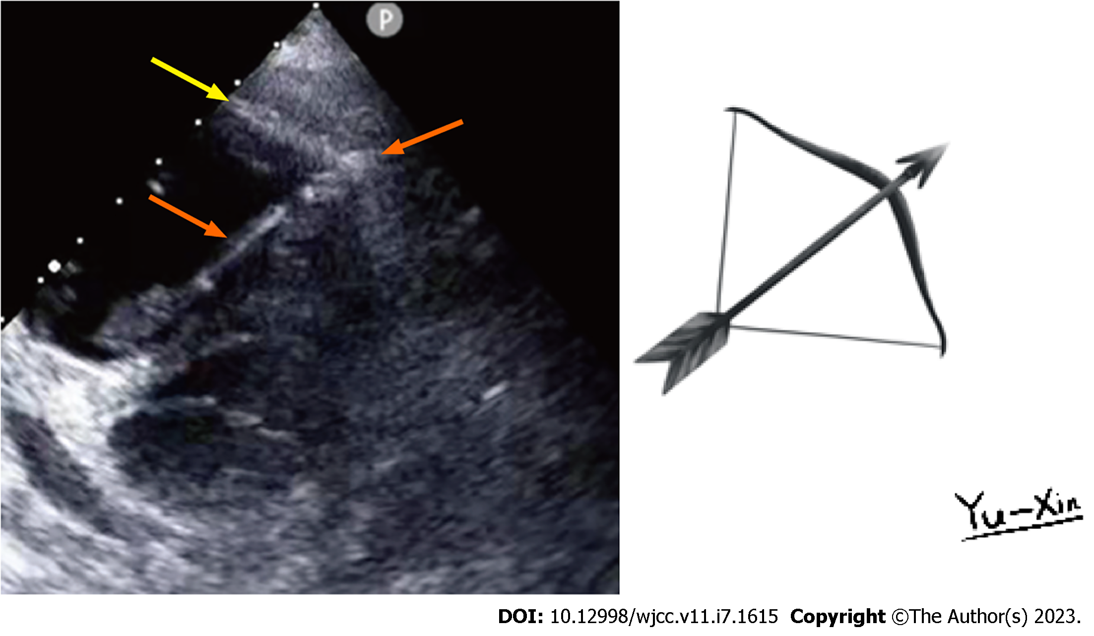
Figure 3 A “bow-and-arrow” sign on point-of-care ultrasound and a sketch of this sign.
The sign consists of the curved outline of the ventricular wall (the bow), the lead shaft (arrow shaft), and the lead tip (arrowhead). The sketch was painted by Yu-Xin Wang.
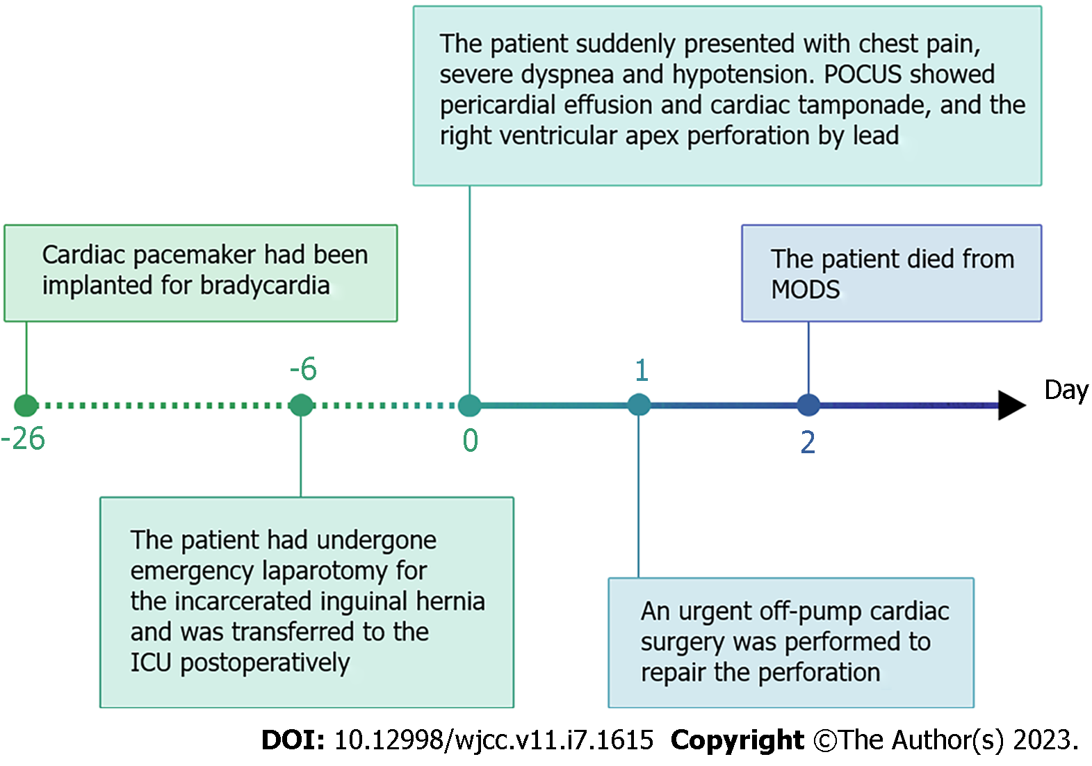
Figure 4 Timeline of the clinical course.
ICU: Intensive care unit; POCUS: Point-of-care ultrasound; MODS: Multiple organ dysfunction syndrome.
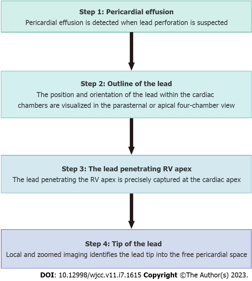
Figure 5 A step-wise approach for rapid diagnosis of lead-induced right ventricular apex perforation by point-of-care ultrasound.
RV: Right ventricle.
- Citation: Chen N, Miao GX, Peng LQ, Li YH, Gu J, He Y, Chen T, Fu XY, Xing ZX. Bow-and-arrow sign on point-of-care ultrasound for diagnosis of pacemaker lead-induced heart perforation: A case report and literature review. World J Clin Cases 2023; 11(7): 1615-1626
- URL: https://www.wjgnet.com/2307-8960/full/v11/i7/1615.htm
- DOI: https://dx.doi.org/10.12998/wjcc.v11.i7.1615


