Copyright
©The Author(s) 2023.
World J Clin Cases. Feb 26, 2023; 11(6): 1426-1433
Published online Feb 26, 2023. doi: 10.12998/wjcc.v11.i6.1426
Published online Feb 26, 2023. doi: 10.12998/wjcc.v11.i6.1426
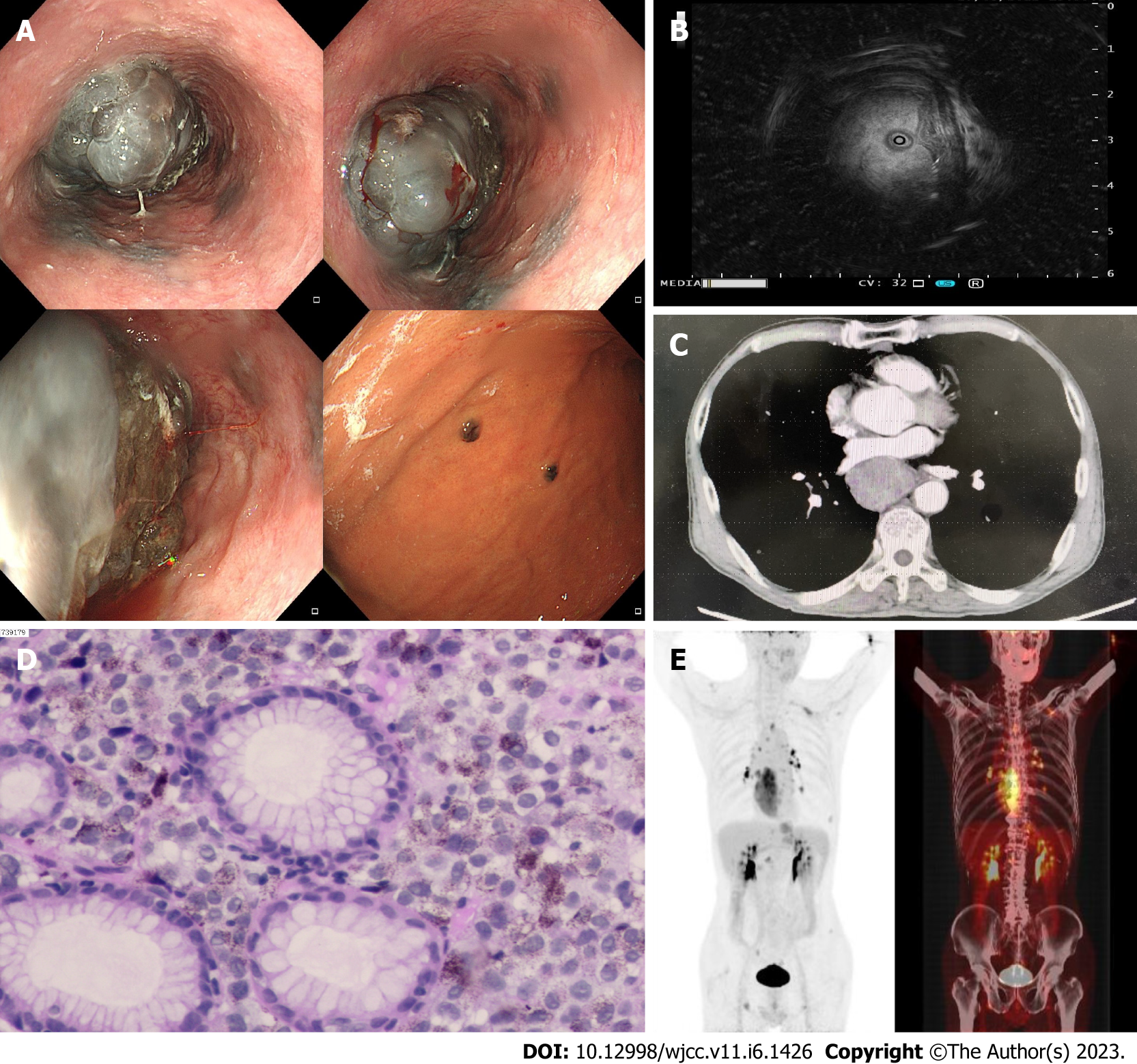
Figure 1 Imaging and pathological examinations of the patient with primary malignant melanoma of the esophagus.
A: Gastroscopy revealed in the esophagus (30-40 cm from the incisor) a bulging mass with blackish color, rough surface, seeming erosion in the middle, and easy bleeding on contact. The gastric fundus and mucosa of the gastric body were scattered with multiple, flat, blackish elevated lesions of approximately 0.4-0.6 cm in size; B: Ultrasound endoscopy demonstrated that the main lesion had mixed echogenicity, predominantly hypoechogenicity, and involved the entire wall of the duct, with enlarged lymph nodes visible outside the wall; C: Chest contrast-enhanced computed tomography showed a soft tissue mass in the middle and lower esophagus; D: Pathology showed uniform and consistent heterogeneous cell infiltration in the intrinsic layer; the cells were small and round with clear boundaries; some of the cytoplasm carried pigment granules; and the nucleus was clearly chromatin fine; E: Whole-body positron emission tomography/computed tomography showed hypermetabolic occupancy in the lower and middle esophagus, multiple hypermetabolic lymph nodes throughout the body, and multiple hypermetabolic foci in bone.
- Citation: Wang QQ, Li YM, Qin G, Liu F, Xu YY. Primary malignant melanoma of the esophagus: A case report. World J Clin Cases 2023; 11(6): 1426-1433
- URL: https://www.wjgnet.com/2307-8960/full/v11/i6/1426.htm
- DOI: https://dx.doi.org/10.12998/wjcc.v11.i6.1426


