Copyright
©The Author(s) 2023.
World J Clin Cases. Feb 26, 2023; 11(6): 1393-1402
Published online Feb 26, 2023. doi: 10.12998/wjcc.v11.i6.1393
Published online Feb 26, 2023. doi: 10.12998/wjcc.v11.i6.1393
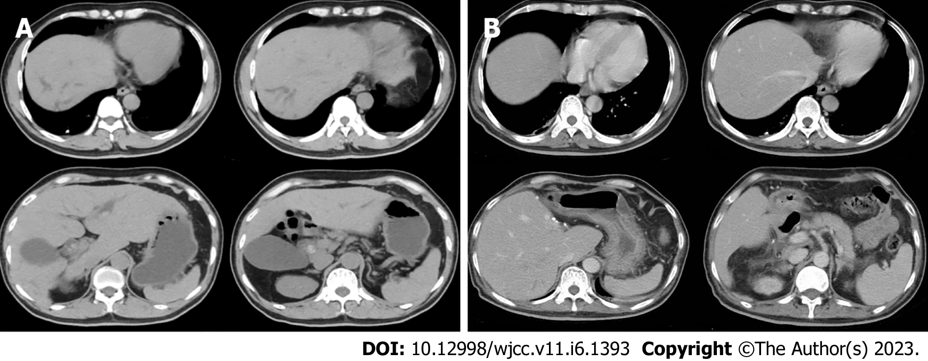
Figure 1 Computed tomography image.
A: The preoperative computed tomography (CT) image of the upper abdomen of the patient: Normal liver parenchyma with dilated intrahepatic as well as common bile duct (about 18 mm) and enlarged gallbladder without wall-thickening; B: Postoperative CT image of the upper abdomen: the enlarged and deformed remnant liver with dilated intrahepatic bile duct in the upper segment of the right posterior; the portal vein is widened, the widest diameter is about 1.4 cm. The spleen was normal in size and shape.
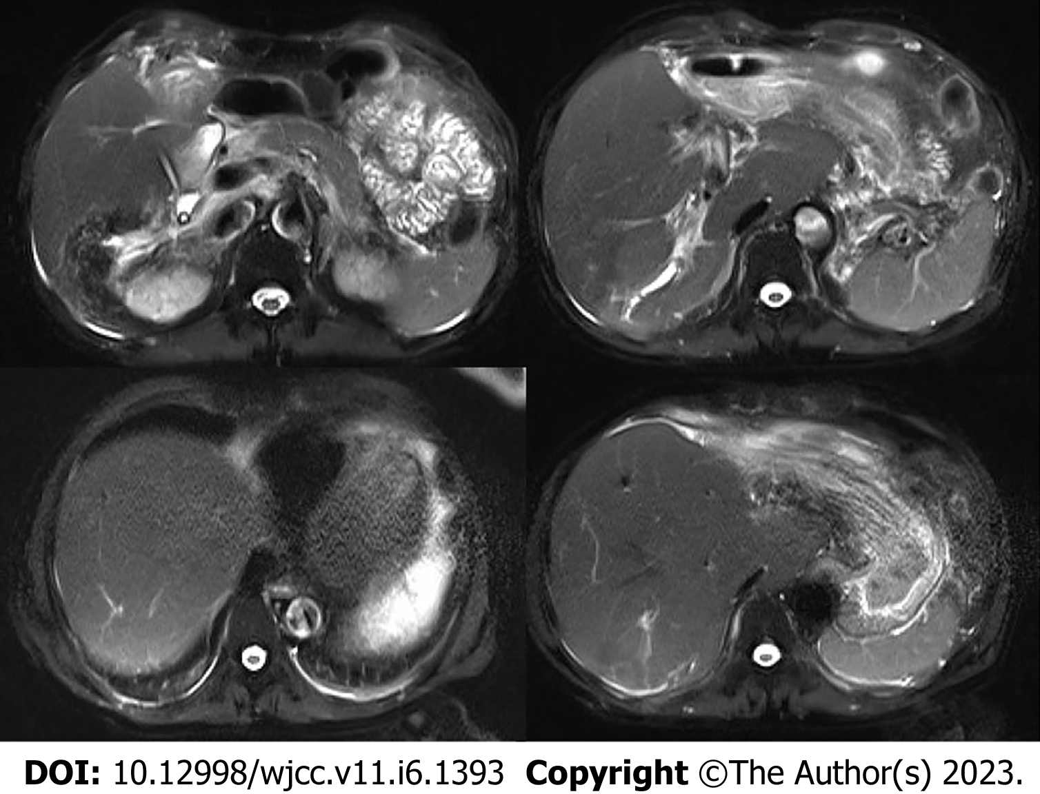
Figure 2 Postoperative magnetic resonance imaging of the upper abdomen.
Enlarged and deformed liver with uneven signals in the liver parenchyma and dilated intrahepatic bile duct in the upper right posterior lobe of the liver. Multiple nodular short T2 signal in the dilated and rigid bile duct with increased T2 weighted imaging signal in the surrounding liver parenchyma. The portal vein is widened with a size of 1.4 cm.
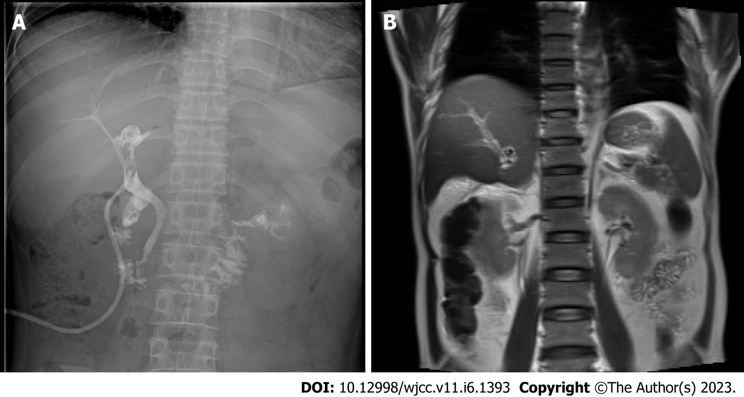
Figure 3 T-tube and magnetic resonance cholangiopancreatography images.
A: T-tube angiography: the left and right hepatic ducts were not visualized, the bile duct was rigid, and the lower end of the common bile duct was unobstructed; B: Magnetic resonance cholangiopancreatography: The intrahepatic bile duct in the upper right posterior lobe of the liver was dilated, and multiple nodular short T2 signals were seen in the lumen. Gallbladder not shown.
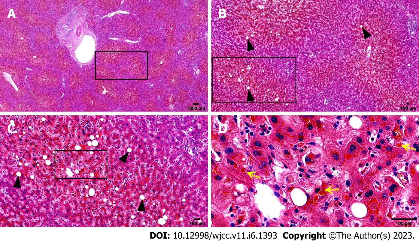
Figure 4 Histopathological biopsy of the liver.
The portal area was enlarged to varying degrees, with hyperplasia of fibrous tissue, infiltration of a moderate amount of lymphocytes and a small number of plasma cells, minor bile duct damage, fibrosis around a small number of small bile ducts, and mild bile duct hyperplasia, mild edema of hepatocytes, a few balloon-like degenerated hepatocytes, a few glycogenuclear hepatocytes, bulicular and microbulicular steatosis of hepatocytes (hepatic steatosis cells < 5%), irregular distribution, rare punctate necrosis. Some hepatocytes cholestatic pigment granules can be seen, and some bile ducts are dilated and cholestatic. Yellow arrow: Cholestatic pigment particles; Black triangle: Hepatocyte adipose change. A: Hematoxylin-eosin (HE) (× 1); B: HE (× 4); C: HE (× 10); D: HE (× 40).
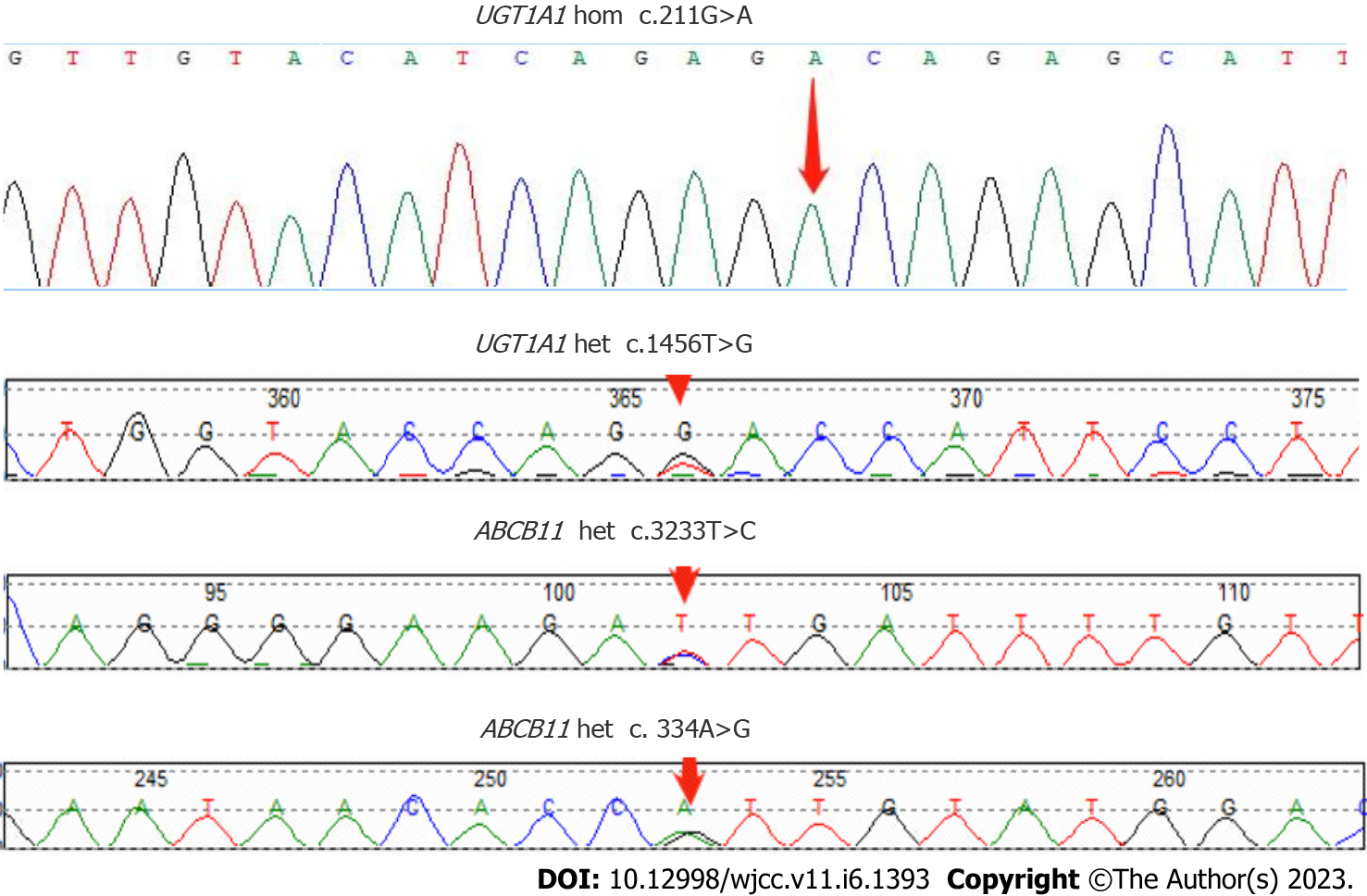
Figure 5 Sanger sequencing map of the patient's ABCB11 and UGT1A1 genes with designated mutation sites.
- Citation: Jiang JL, Liu X, Pan ZQ, Jiang XL, Shi JH, Chen Y, Yi Y, Zhong WW, Liu KY, He YH. Postoperative jaundice related to UGT1A1 and ABCB11 gene mutations: A case report and literature review. World J Clin Cases 2023; 11(6): 1393-1402
- URL: https://www.wjgnet.com/2307-8960/full/v11/i6/1393.htm
- DOI: https://dx.doi.org/10.12998/wjcc.v11.i6.1393


