Copyright
©The Author(s) 2023.
World J Clin Cases. Feb 26, 2023; 11(6): 1365-1371
Published online Feb 26, 2023. doi: 10.12998/wjcc.v11.i6.1365
Published online Feb 26, 2023. doi: 10.12998/wjcc.v11.i6.1365
Figure 1 Transvaginal ultrasonographic examination of the uterine cavity and fallopian tube.
A and B: Transvaginal sonography indicates a mixed lesion (A–B) with a non-homogeneous echo (yellow arrow) in the uterine cavity and a solid cyst (yellow arrow) (C) of the left adnexa; both lesions are accompanied by a rich blood flow. A low resistance index (RI, 0.78) and high arterial spectrum (Vmax, 26 cm/s) of the left adnexal mass (yellow arrow) are also detected.
Figure 2 Fallopian tube endometrioid adenocarcinoma arising from endometriosis synchronized with endometrial endometrioid adenocarcinoma.
A: A distorted and thickened left hydrosalpinx (50.0 mm × 40.0 mm) with a blocked end (yellow arrow) was seen during laparoscopic exploration; B: Diffuse, cauliflower, and fish-like exogenous cancerous lesions (yellow arrow) were filled with feculent liquid in the left fallopian tube; C and D: The endometrioid adenocarcinoma in the uterus was grade 1, with a myometrial invasion of less than 50% (Hematoxylin-eosin Staining, HE, ×100, yellow arrow); E: Endometrioid adenocarcinoma of the fallopian tube (HE, ×100, yellow arrow); F: The endometriotic area in the mucosa of the left fallopian tube (HE, ×100, yellow arrow); G and H: The transitional area from endometriosis to atypical hyperplasia in the left fallopian tube (G, HE, ×100; H, HE, ×200, yellow arrow).
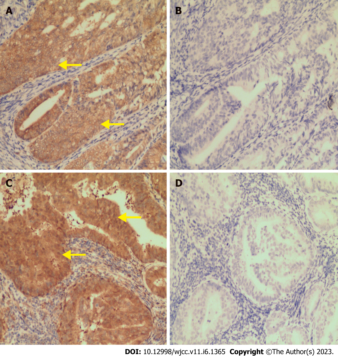
Figure 3 PTEN and PAX2 expression patterns in endometrial and fallopian tube endometrioid adenocarcinoma.
Both endometrial- and fallopian-tube endometrioid adenocarcinoma showed the same model for positive expression of PTEN (A and C, IHC, ×100, yellow arrow) and loss of expression of PAX2 (B and D, IHC, ×100).
- Citation: Feng JY, Jiang QP, He H. Endometriosis-associated endometrioid adenocarcinoma of the fallopian tube synchronized with endometrial adenocarcinoma: A case report. World J Clin Cases 2023; 11(6): 1365-1371
- URL: https://www.wjgnet.com/2307-8960/full/v11/i6/1365.htm
- DOI: https://dx.doi.org/10.12998/wjcc.v11.i6.1365


