Copyright
©The Author(s) 2023.
World J Clin Cases. Feb 16, 2023; 11(5): 1206-1216
Published online Feb 16, 2023. doi: 10.12998/wjcc.v11.i5.1206
Published online Feb 16, 2023. doi: 10.12998/wjcc.v11.i5.1206
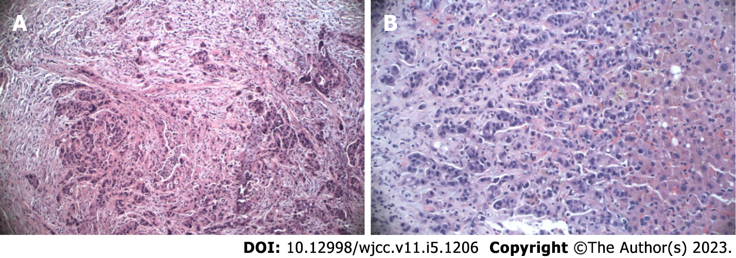
Figure 1 Histopathology.
A: Histopathological analysis. Hematoxylin and eosin staining (× 200) revealed stage IV pancreatic ductal adenocarcinoma of the head; B: Histopathological examination. Hematoxylin and eosin staining (× 200) revealed liver metastasis.
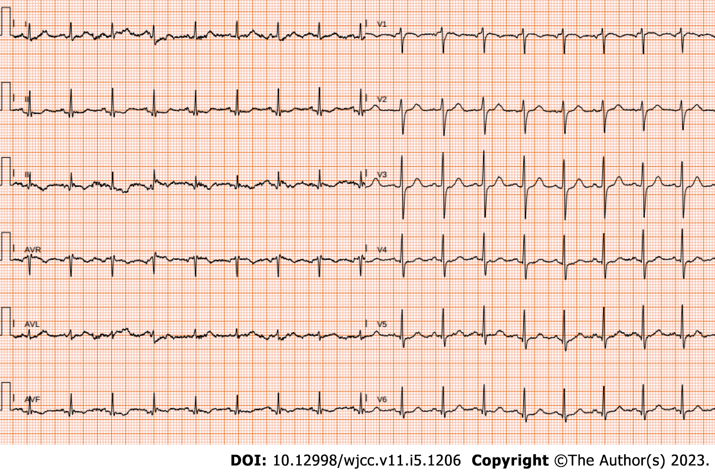
Figure 2 Baseline electrocardiogram.
Sinus tachycardia and biphasic T waves in the inferior leads were observed. The patient’s heart rate was 103 beats/min at rest.
Figure 3 Computed tomography.
A: The axial view revealed partial occlusion of the right branch of the pulmonary artery (arrow); B: The axial view revealed partial occlusion of the left branch of the pulmonary artery (arrow).
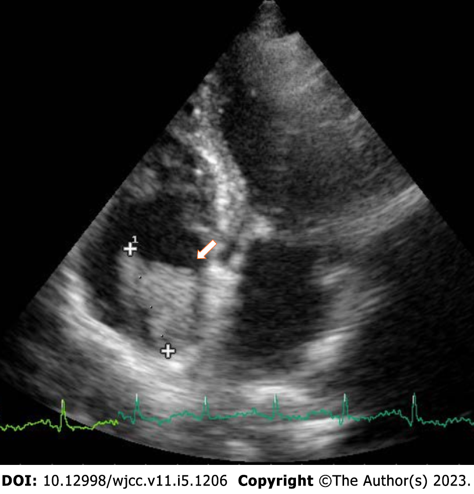
Figure 4 Transthoracic echocardiography.
The apical four-chamber view revealed a large solid isoechoic/hyperechoic mass in the right atrium (2 cm × 3 cm) (arrow).
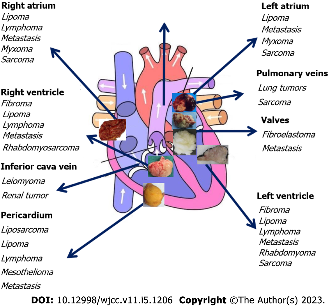
Figure 5 Typical localization of cardiac tumors.
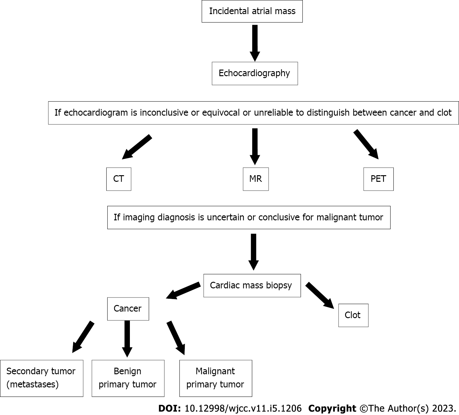
Figure 6 Diagnostic non-invasive algorithm to differentiate thrombi from cancers.
CT: Computed tomography; MR: Magnetic resonance; PET: Positron emission tomography.
- Citation: Fioretti AM, Leopizzi T, La Forgia D, Scicchitano P, Oreste D, Fanizzi A, Massafra R, Oliva S. Incidental right atrial mass in a patient with secondary pancreatic cancer: A case report and review of literature. World J Clin Cases 2023; 11(5): 1206-1216
- URL: https://www.wjgnet.com/2307-8960/full/v11/i5/1206.htm
- DOI: https://dx.doi.org/10.12998/wjcc.v11.i5.1206


