Copyright
©The Author(s) 2023.
World J Clin Cases. Feb 16, 2023; 11(5): 1198-1205
Published online Feb 16, 2023. doi: 10.12998/wjcc.v11.i5.1198
Published online Feb 16, 2023. doi: 10.12998/wjcc.v11.i5.1198
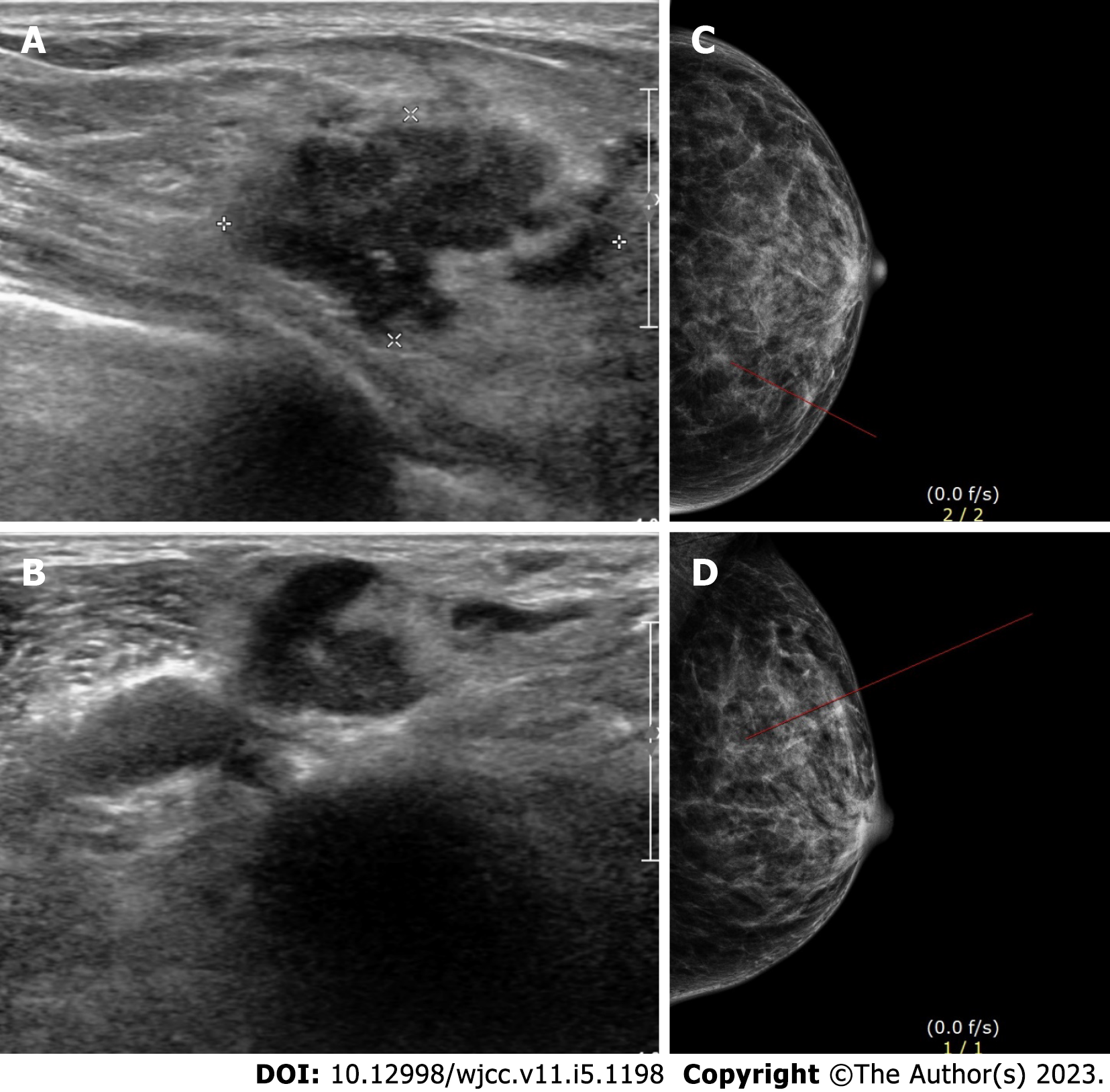
Figure 1 Imaging of the left breast lump.
A: Ultrasonography of the left breast; B: Ultrasonography of the left breast; C: Craniocaudal view of mammography of the left breast; D: Mediolateral oblique view of mammography of the left breast.
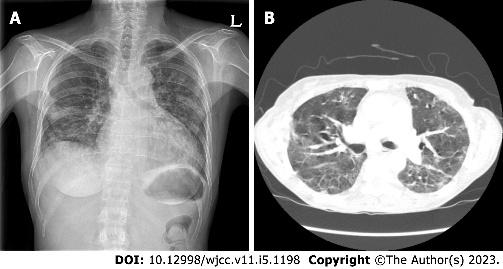
Figure 2 Imaging of the chest.
A: Chest radiograph showing multiple patch opacities in both lungs; B: Computed tomography showing multifocal patch ground-glass opacity consolidation in both lungs.
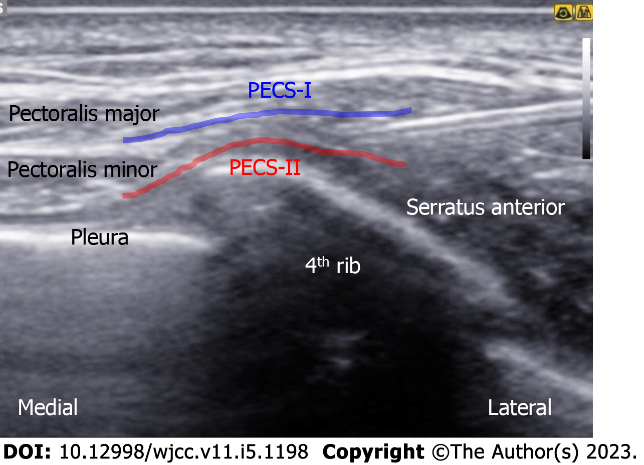
Figure 3 Ultrasonography of pectoral nerve block type II.
PECS-I: Pectoral nerve block type I; PECS-II: Pectoral nerve block type II.
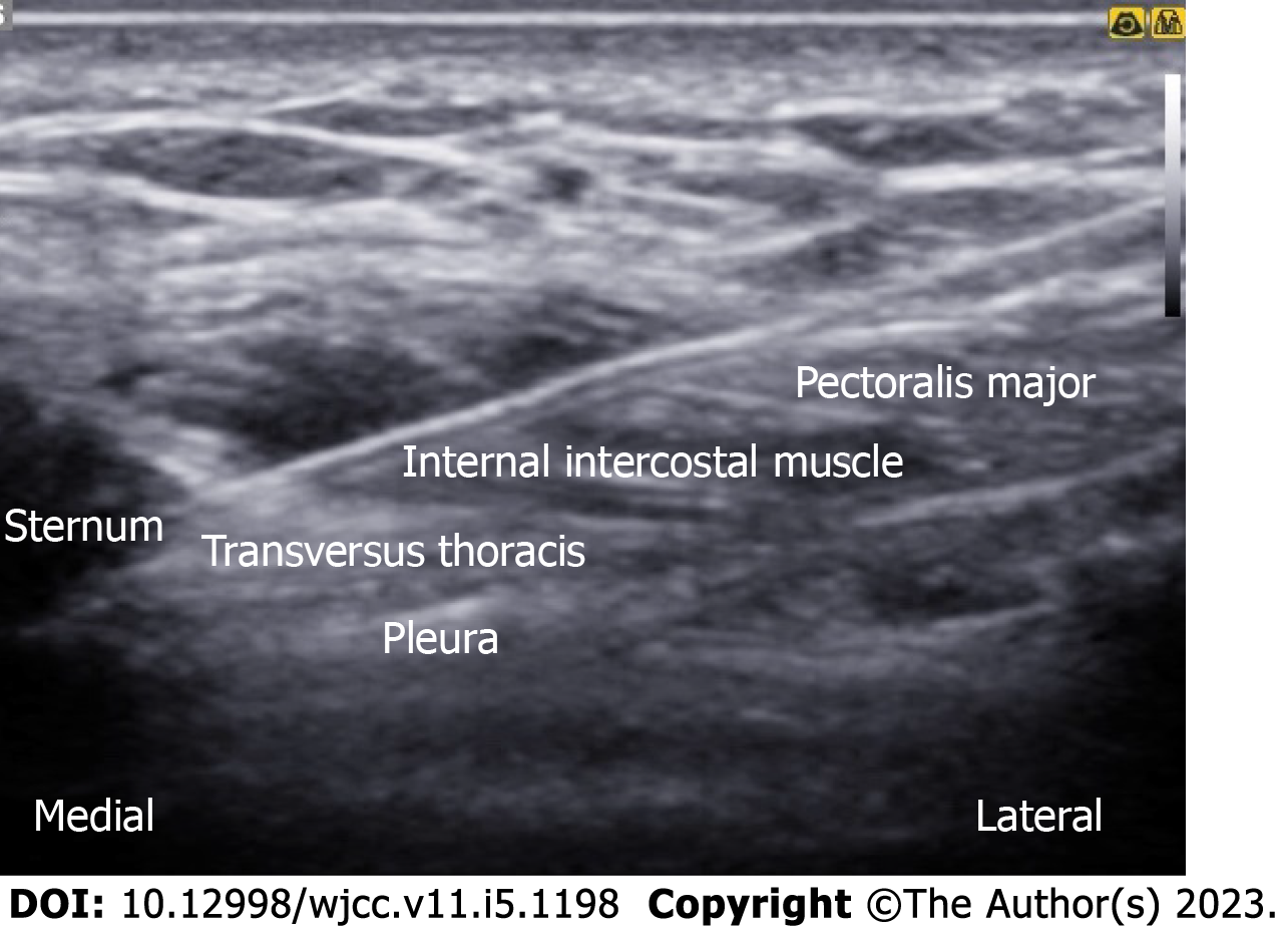
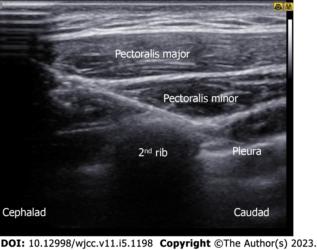
Figure 4 Ultrasonography of parasternal nerve block.
Figure 5 Ultrasonography of intercostobrachial nerve block.
- Citation: Jin Y, Lee S, Kim D, Hur J, Eom W. Combinations of nerve blocks in surgery for post COVID-19 pulmonary sequelae patient: A case report and review of literature. World J Clin Cases 2023; 11(5): 1198-1205
- URL: https://www.wjgnet.com/2307-8960/full/v11/i5/1198.htm
- DOI: https://dx.doi.org/10.12998/wjcc.v11.i5.1198


