Copyright
©The Author(s) 2023.
World J Clin Cases. Feb 16, 2023; 11(5): 1175-1181
Published online Feb 16, 2023. doi: 10.12998/wjcc.v11.i5.1175
Published online Feb 16, 2023. doi: 10.12998/wjcc.v11.i5.1175
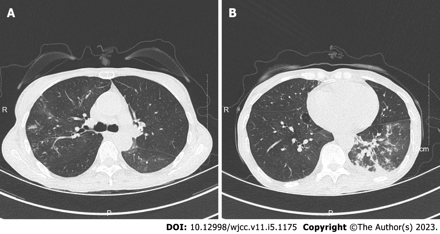
Figure 1 Computerized tomography showed multiple patchy, nodular and strip high density shadows in both lungs.
A: Showed multiple patchy, nodular and strip-shaped high-density shadows in the upper lobe of the right lung; B: Showed multiple patchy, nodular and strip-shaped high-density shadows in the upper lobe of the lower lobe of the left lung, multiple sputum thrombi in the bronchus of the lower lobe of the left lung.
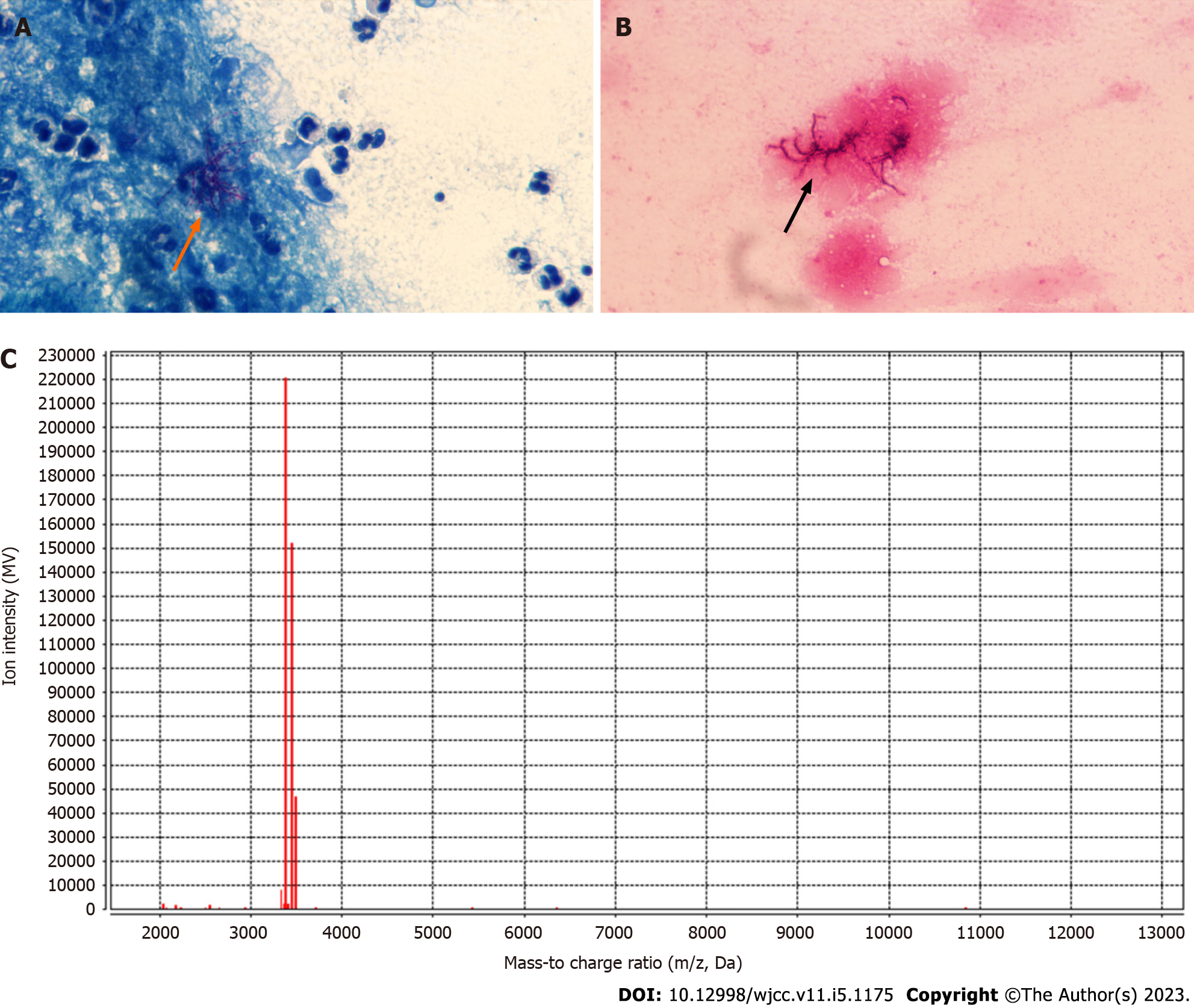
Figure 2 Pathogenic examination.
A: Positive acid-fast bifurcating filaments were observed in bronchoalveolar lavage fluid of the patient under oil microscope (×1000 magnification); B: Branching gram-positive rods were observed in bronchoalveolar lavage fluid of the patient under oil microscope (×1000 magnification); C: matrix-assisted laser desorption ionization-time of flight mass spectrometry confirm it was Nocardia cyriacigeorgica.
- Citation: Hong X, Ji YQ, Chen MY, Gou XY, Ge YM. Nocardia cyriacigeorgica infection in a patient with repeated fever and CD4+ T cell deficiency: A case report. World J Clin Cases 2023; 11(5): 1175-1181
- URL: https://www.wjgnet.com/2307-8960/full/v11/i5/1175.htm
- DOI: https://dx.doi.org/10.12998/wjcc.v11.i5.1175


