Copyright
©The Author(s) 2023.
World J Clin Cases. Feb 16, 2023; 11(5): 1158-1164
Published online Feb 16, 2023. doi: 10.12998/wjcc.v11.i5.1158
Published online Feb 16, 2023. doi: 10.12998/wjcc.v11.i5.1158
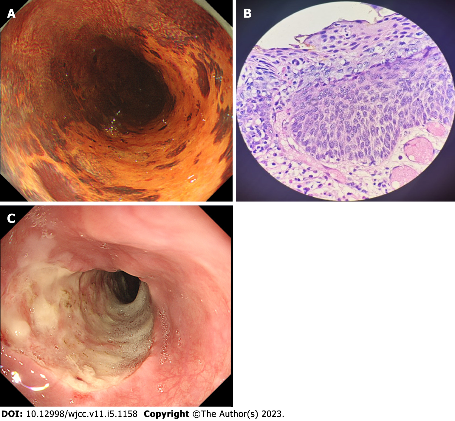
Figure 1 Histology of the esophageal mucosa before endoscopic submucosal dissection and the wound surface after treatment.
A: Iodine staining showing extension of the target lesion to almost two-thirds of the esophageal circumference, including a small non-stained area; B: Pathological examination of biopsy specimen showing severe squamous cell dysplasia; C: En-bloc lesion resection was performed five days after endoscopic submucosal dissection; the formation of ulcers can be seen on the wound.
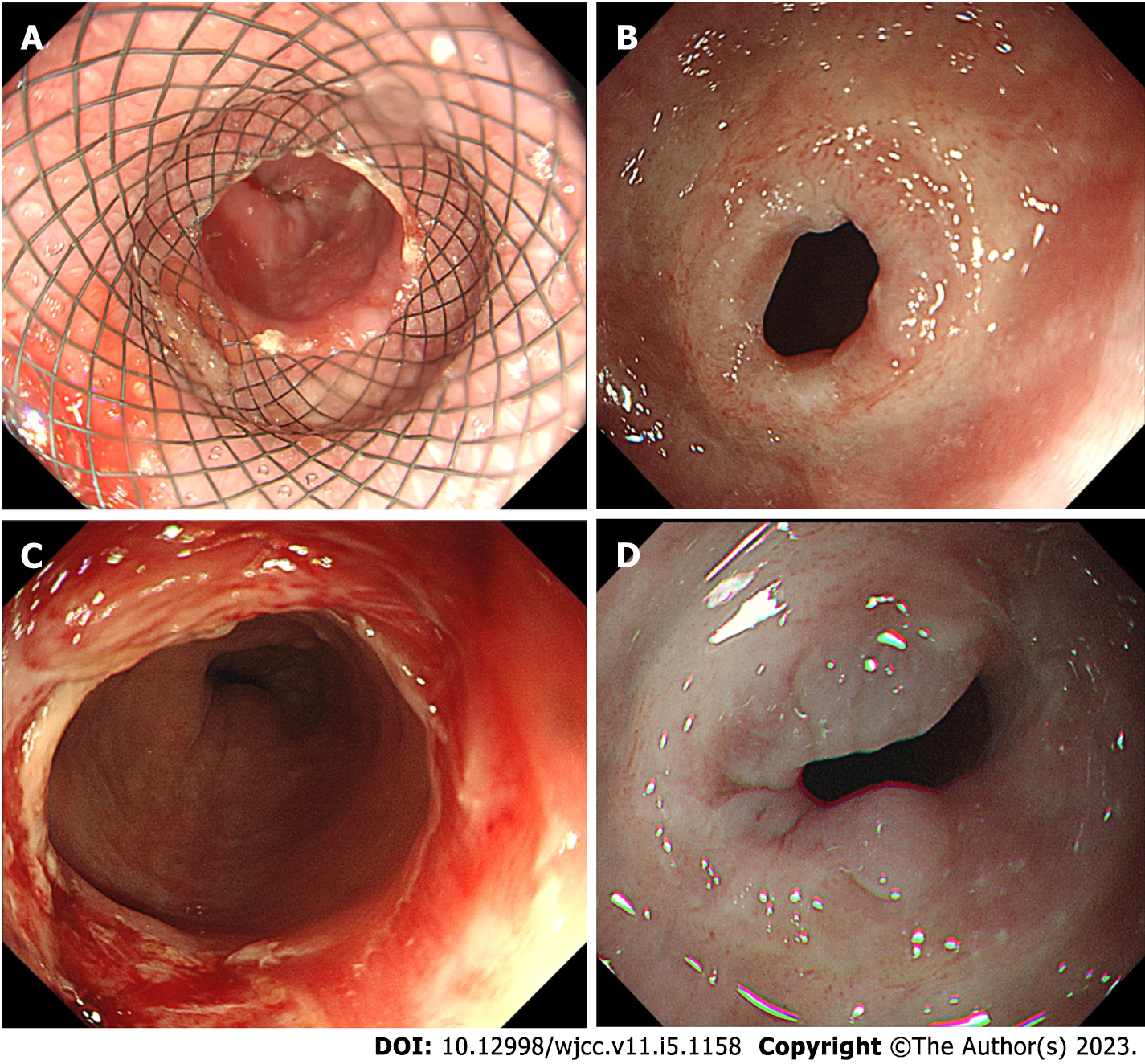
Figure 2 Stent removal and stricture formation; Stricture occurred after the removal of the stent and endoscopic dilatation was performed but proved ineffective.
A: An esophageal stent was implanted five days after endoscopic submucosal dissection; B: Esophageal stricture 37 cm from the incisors; C: Bougie dilation was performed at the stenosis site; D: The stricture was still apparent even after three expansion treatments.
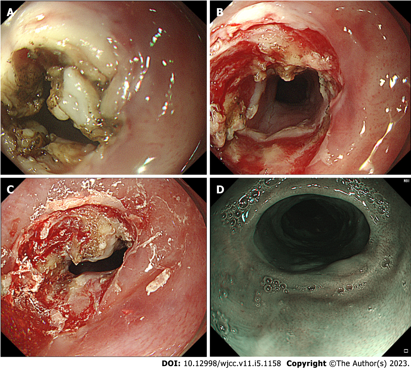
Figure 3 Combination of radial incision and cutting, dilation, and steroid injection treatment.
A: Radial incision and cutting of the mucosa and submucosal fibrous scar in four places; B: Expansion treatment to fully open the stricture ring; C: The steroid triamcinolone acetonide was injected into the surface of the stricture and surrounding mucosa; D: Three months post-treatment, the gastroscope was able to pass without any resistance.
- Citation: Pu WF, Zhang T, Du ZH. Combined treatment of refractory benign stricture after esophageal endoscopic mucosal dissection: A case report. World J Clin Cases 2023; 11(5): 1158-1164
- URL: https://www.wjgnet.com/2307-8960/full/v11/i5/1158.htm
- DOI: https://dx.doi.org/10.12998/wjcc.v11.i5.1158


