Copyright
©The Author(s) 2023.
World J Clin Cases. Feb 16, 2023; 11(5): 1152-1157
Published online Feb 16, 2023. doi: 10.12998/wjcc.v11.i5.1152
Published online Feb 16, 2023. doi: 10.12998/wjcc.v11.i5.1152
Figure 1 Thyroid ultrasonography revealed a lobulated solid hypoechoic nodule with unclear boundaries and little blood flow signal.
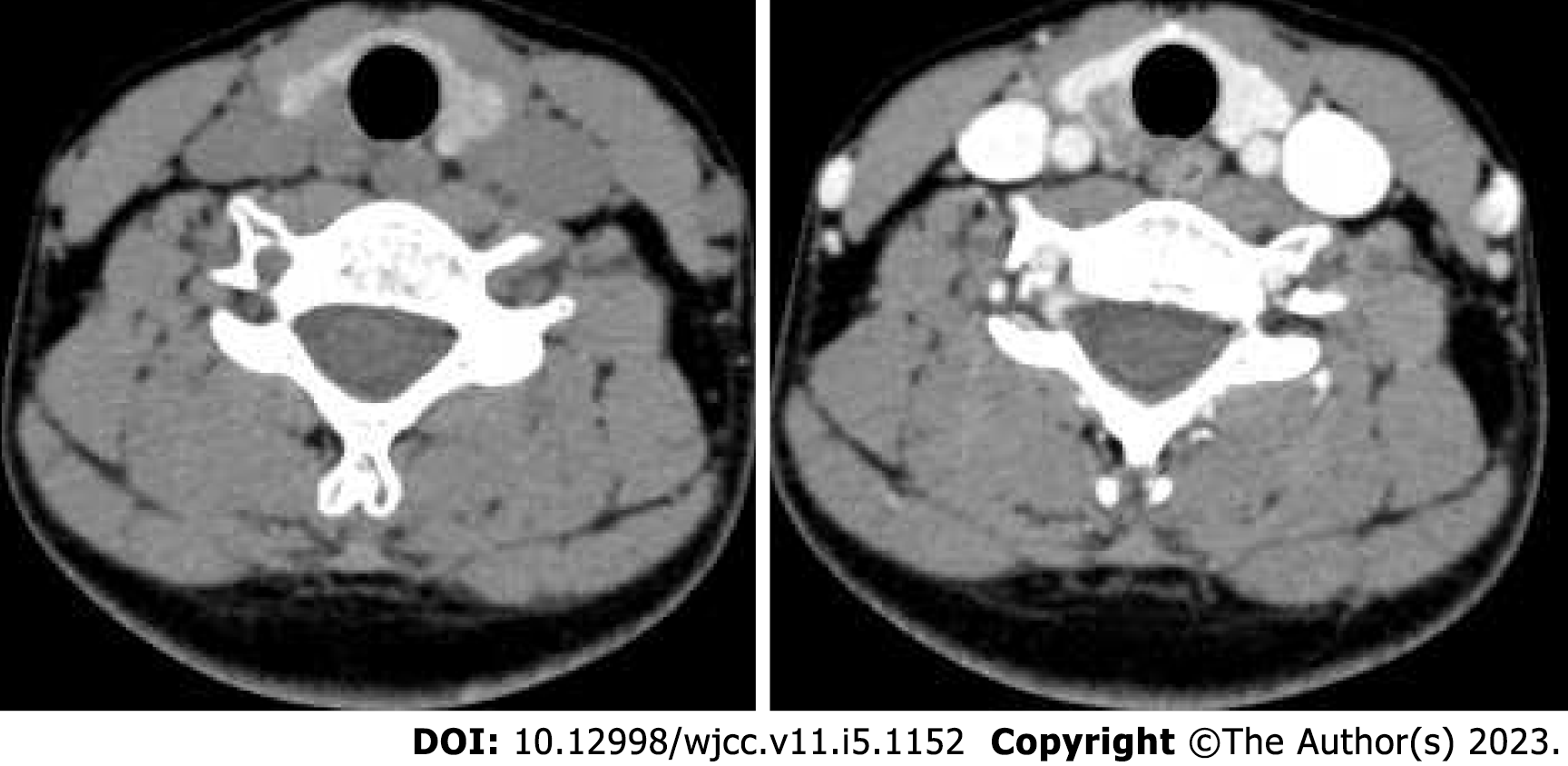
Figure 2 Contrast-enhanced computed tomography of the neck showed patchy hypodense foci in the right thyroid gland, with less enhancement than the thyroid gland itself.
- Citation: Shi JJ, Peng Y, Zhang Y, Zhou L, Pan G. Langerhans cell histiocytosis misdiagnosed as thyroid malignancy: A case report. World J Clin Cases 2023; 11(5): 1152-1157
- URL: https://www.wjgnet.com/2307-8960/full/v11/i5/1152.htm
- DOI: https://dx.doi.org/10.12998/wjcc.v11.i5.1152


