Copyright
©The Author(s) 2023.
World J Clin Cases. Feb 16, 2023; 11(5): 1144-1151
Published online Feb 16, 2023. doi: 10.12998/wjcc.v11.i5.1144
Published online Feb 16, 2023. doi: 10.12998/wjcc.v11.i5.1144
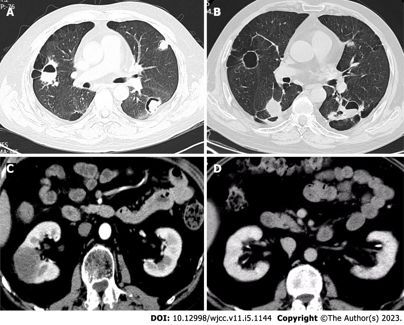
Figure 1 Computed tomography.
A: On pulmonary computed tomography (CT), multiple patchy shadows, nodular higher-density shadows, multiple cavities, heterogeneous wall thickness, and air crescent sign and flat lipid in some lesions were observed (August 13, 2018); B: On pulmonary CT, there were new and larger bilateral pulmonary nodules (June 12, 2020); C: Renal CT was performed and revealed a space-occupying lesion in the right middle kidney, which was larger than before (June 15, 2021); D: The right middle kidney-occupying lesion was significantly absorbed after tocilizumab treatment (August 20, 2021).
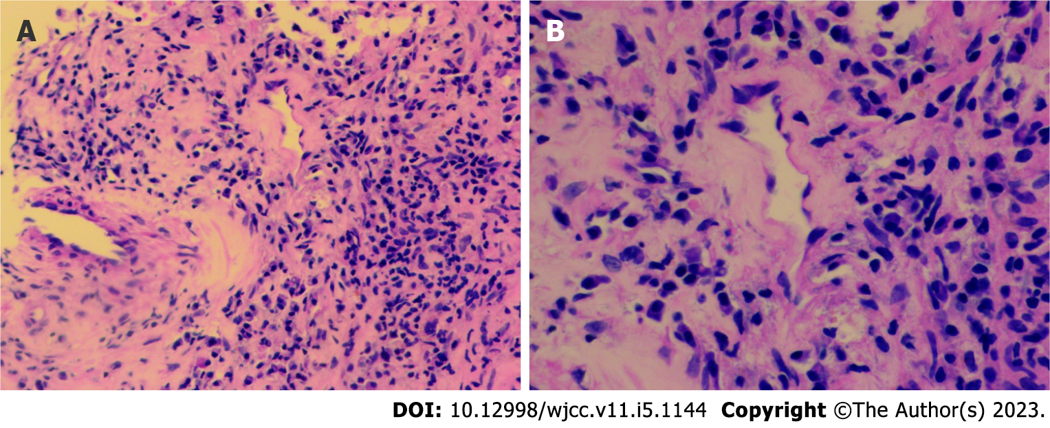
Figure 2 Puncture pathology.
A and B: Puncture pathology of the space-occupying lesion revealed infiltration of tissue cells, plasma cells, and lymphocytes (A: magnification × 200; B: Magnification × 400).
Figure 3 Erythrocyte sedimentation rate and the levels of interleukin-6 and proteinase 3-antineutrophil cytoplasmic antibody.
A: Erythrocyte sedimentation rate; B: Interleukin-6; C: Proteinase 3-antineutrophil cytoplasmic antibody. ESR: Erythrocyte sedimentation rate; IL-6: Interleukin-6; CTX: Cyclophosphamide; TW: Tripterygium wilfordii; MMF: Mycophenolate mofetil; TCZ: Tocilizumab.
- Citation: Tang PF, Xu LC, Hong WT, Shi HY. Successful treatment of granulomatosis with polyangiitis using tocilizumab combined with glucocorticoids: A case report. World J Clin Cases 2023; 11(5): 1144-1151
- URL: https://www.wjgnet.com/2307-8960/full/v11/i5/1144.htm
- DOI: https://dx.doi.org/10.12998/wjcc.v11.i5.1144


