Copyright
©The Author(s) 2023.
World J Clin Cases. Feb 16, 2023; 11(5): 1137-1143
Published online Feb 16, 2023. doi: 10.12998/wjcc.v11.i5.1137
Published online Feb 16, 2023. doi: 10.12998/wjcc.v11.i5.1137
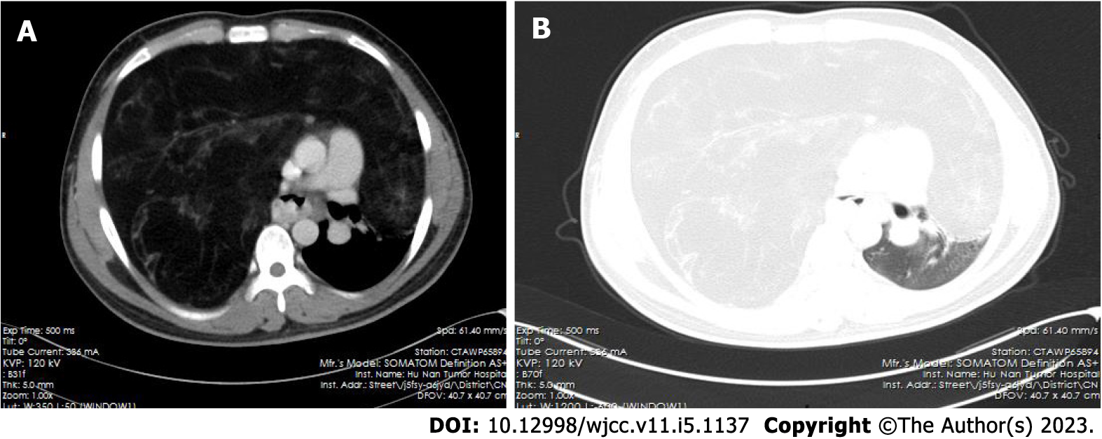
Figure 1 High-resolution chest contrast-enhanced computed tomography.
A: Mediastinal window; B: Lung window. A large fat-containing mass in the anterior mediastinum occupied almost the entire right thoracic cavity and part of the left thoracic cavity with a clear boundary, resulting in lung collapse and mediastinal shift.
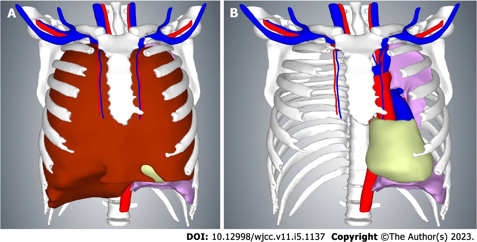
Figure 2 Three-dimensional reconstruction of the chest based on computed tomography images.
A: The anterior mediastinal mass is visible; B: The anterior mediastinal mass is hidden. The volume of the mass as calculated by the three-dimensional reconstruction was 7052 mL.
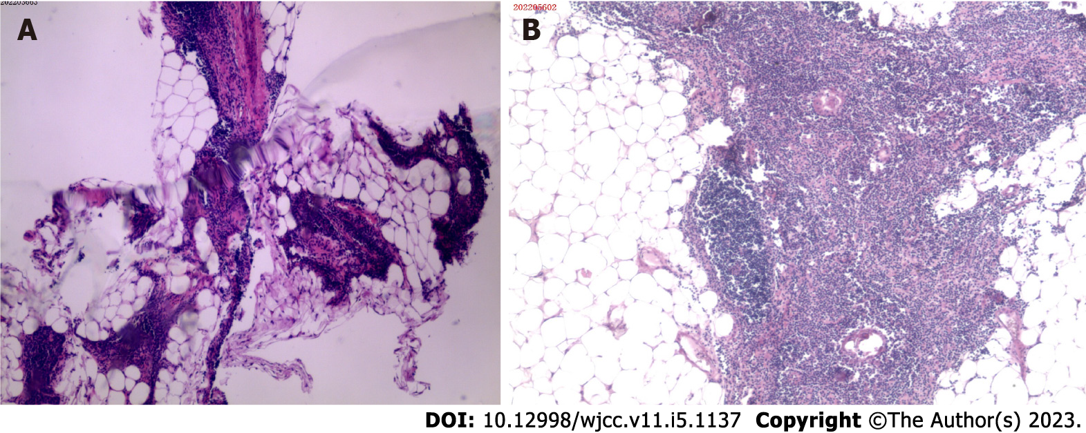
Figure 3 Histopathological results.
A: Histopathological results of computed tomography-guided percutaneous thoracic mass biopsy. Hematoxylin and eosin-stained section showing islands of thymic tissue within mature adipose tissue. Original magnification 40 ×; B: Postoperative histopathological results of the anterior mediastinal mass. Hematoxylin and eosin-stained section showing mature adipose tissue intermixed with septa of atrophic thymus tissue containing lymphocytes and Hassall’s corpuscles. Original magnification 40 ×.
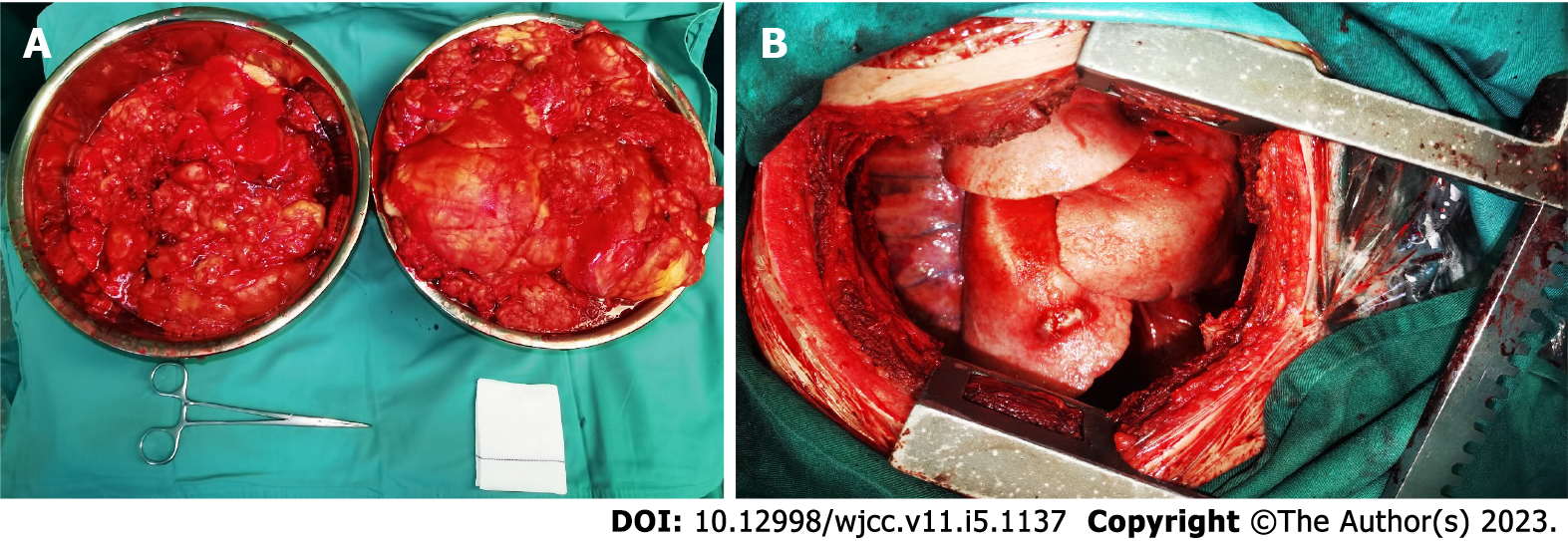
Figure 4 Intraoperative photographs.
A: The tumor was placed in two large containers after piecemeal resection, and the resected tumor weighed 7.5 kg; B: The right thoracic cavity after tumor resection.
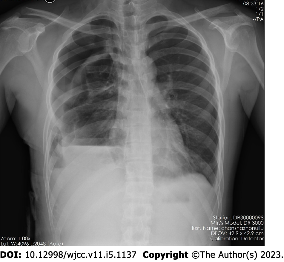
Figure 5 Chest X-ray findings.
The mediastinum had returned to its normal position, the left lung was almost fully inflated, and the right lung was relatively small.
- Citation: Gong LH, Wang WX, Zhou Y, Yang DS, Zhang BH, Wu J. Surgical resection of a giant thymolipoma causing respiratory failure: A case report. World J Clin Cases 2023; 11(5): 1137-1143
- URL: https://www.wjgnet.com/2307-8960/full/v11/i5/1137.htm
- DOI: https://dx.doi.org/10.12998/wjcc.v11.i5.1137


