Copyright
©The Author(s) 2023.
World J Clin Cases. Feb 16, 2023; 11(5): 1122-1128
Published online Feb 16, 2023. doi: 10.12998/wjcc.v11.i5.1122
Published online Feb 16, 2023. doi: 10.12998/wjcc.v11.i5.1122
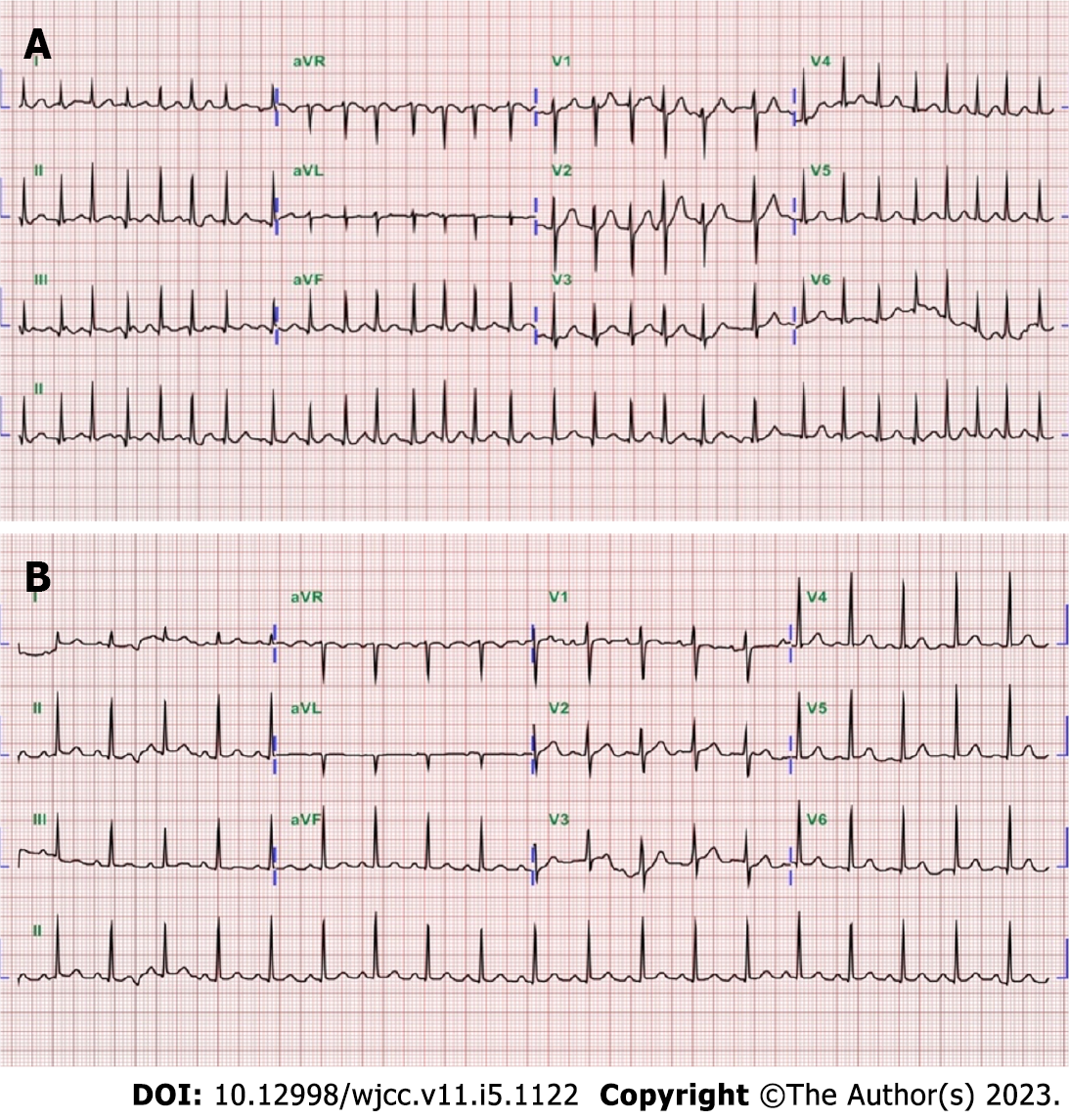
Figure 1 An electrocardiography of cases 1 and 2.
A: Atrial fibrillation with a rapid ventricular response of heart rate 170 beats per minute (case 1); B: Sinus tachycardia and a heart rate of 114 beats per minute (case 2).
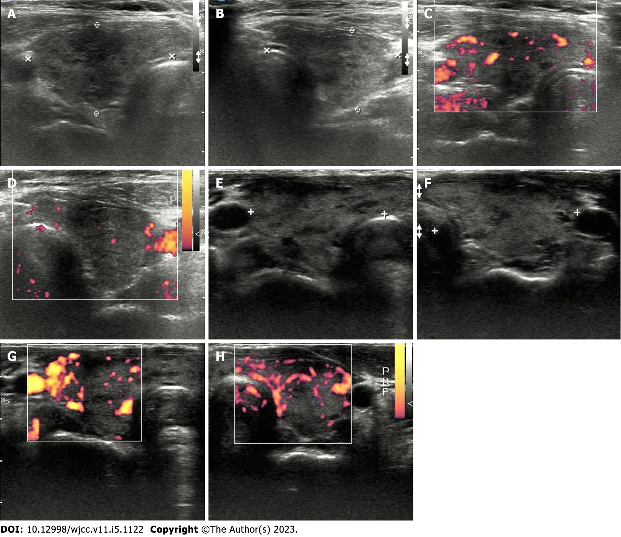
Figure 2 Thyroid ultrasonography of cases 1 and 2.
A: Right thyroid lobe (case 1); B: Left thyroid lobe (case 1); C: Mild increase vascularity in right thyroid lobe (case 1); D: No increase vascularity in left thyroid lobe (case 1); E: Right thyroid lobe (case 2); F: Left thyroid lobe (case 2); G: Increase vascularity in right thyroid lobe (case 2); H: Increase vascularity in left thyroid lobe (case 2).
- Citation: Yan BC, Luo RR. Thyrotoxicosis in patients with a history of Graves’ disease after SARS-CoV-2 vaccination (adenovirus vector vaccine): Two case reports. World J Clin Cases 2023; 11(5): 1122-1128
- URL: https://www.wjgnet.com/2307-8960/full/v11/i5/1122.htm
- DOI: https://dx.doi.org/10.12998/wjcc.v11.i5.1122


