Copyright
©The Author(s) 2023.
World J Clin Cases. Feb 16, 2023; 11(5): 1115-1121
Published online Feb 16, 2023. doi: 10.12998/wjcc.v11.i5.1115
Published online Feb 16, 2023. doi: 10.12998/wjcc.v11.i5.1115
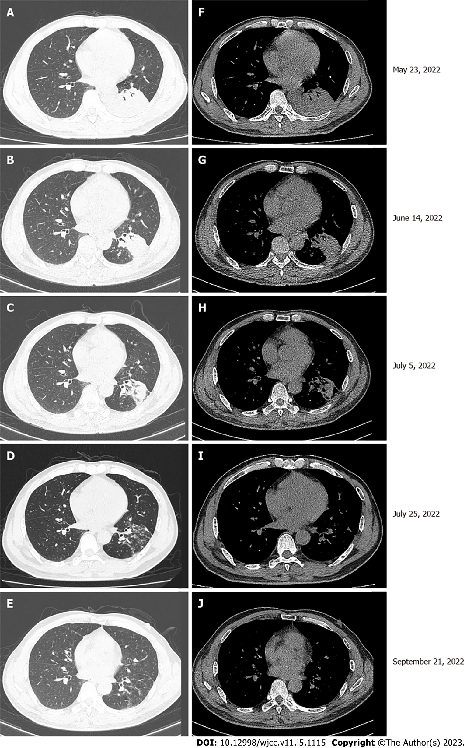
Figure 1 Chest computed tomography.
A and F: A left lower lobe mass with atelectasis was observed; B-D and G-I: After 6 cycles of chemotherapy and immunotherapy, the mass in the left lower lobe gradually decreased in size with treatment; E and J: After 6 cycles of chemotherapy and immunotherapy, the mass in the left lower lobe nearly disappeared by the 6th cycle.
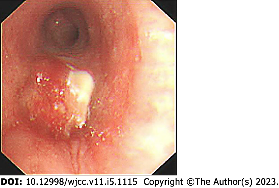
Figure 2
Fiberoptic bronchoscopy revealed a mass protruding from the orifice of the left lower lung inlet, covered with a necrotic white coating and bloody secretions.
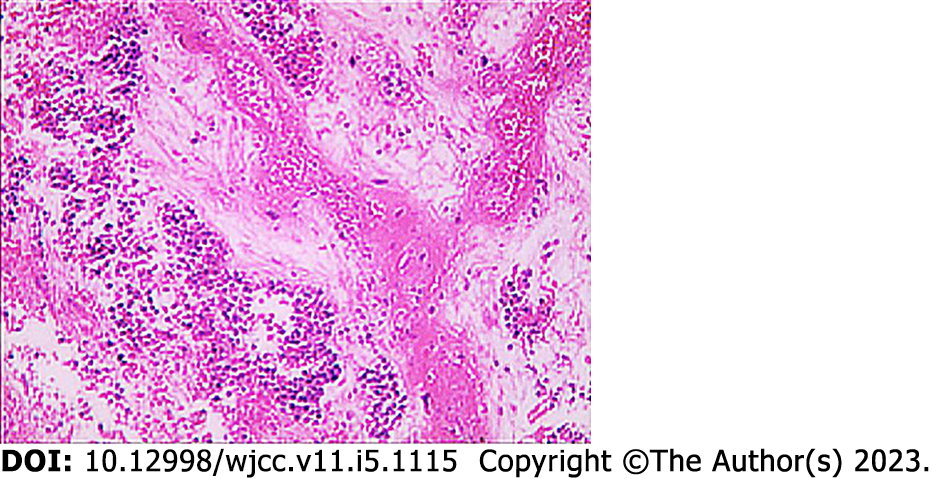
Figure 3 Bronchial mucosal biopsy showed a large amount of necrosis; some cells showed fat spindles, large cells, and nuclear staining, and some cells were small, round and oval with degeneration.
Combined with immunohistochemistry, a diagnosis of neuroendocrine carcinoma and poorly differentiated carcinoma was made.
- Citation: Liu MH, Li YX, Liu Z. Envafolimab combined with chemotherapy in the treatment of combined small cell lung cancer: A case report. World J Clin Cases 2023; 11(5): 1115-1121
- URL: https://www.wjgnet.com/2307-8960/full/v11/i5/1115.htm
- DOI: https://dx.doi.org/10.12998/wjcc.v11.i5.1115


