Copyright
©The Author(s) 2023.
World J Clin Cases. Feb 16, 2023; 11(5): 1106-1114
Published online Feb 16, 2023. doi: 10.12998/wjcc.v11.i5.1106
Published online Feb 16, 2023. doi: 10.12998/wjcc.v11.i5.1106
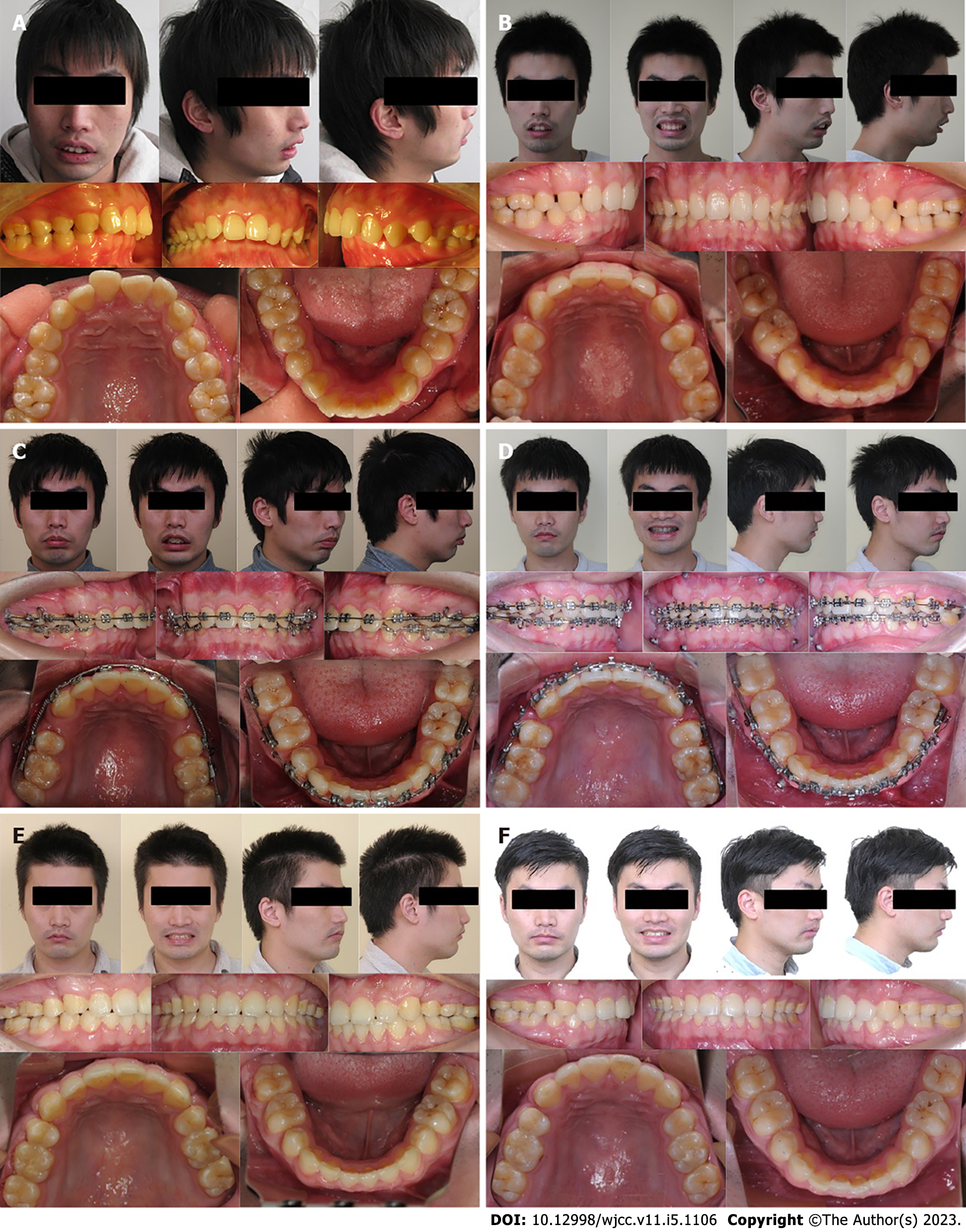
Figure 1 Photographs of the patient's teeth and appearance.
A: Photographs before previous camouflage therapy; B: Pretreatment photographs; C: Pre-surgical photographs; D: Post-surgical photographs; E: Post-treatment photographs; F: 12 mo follow-up photographs.
Figure 2 Dental casts of the patient.
A: Pretreatment dental casts; B: Post-treatment dental casts.
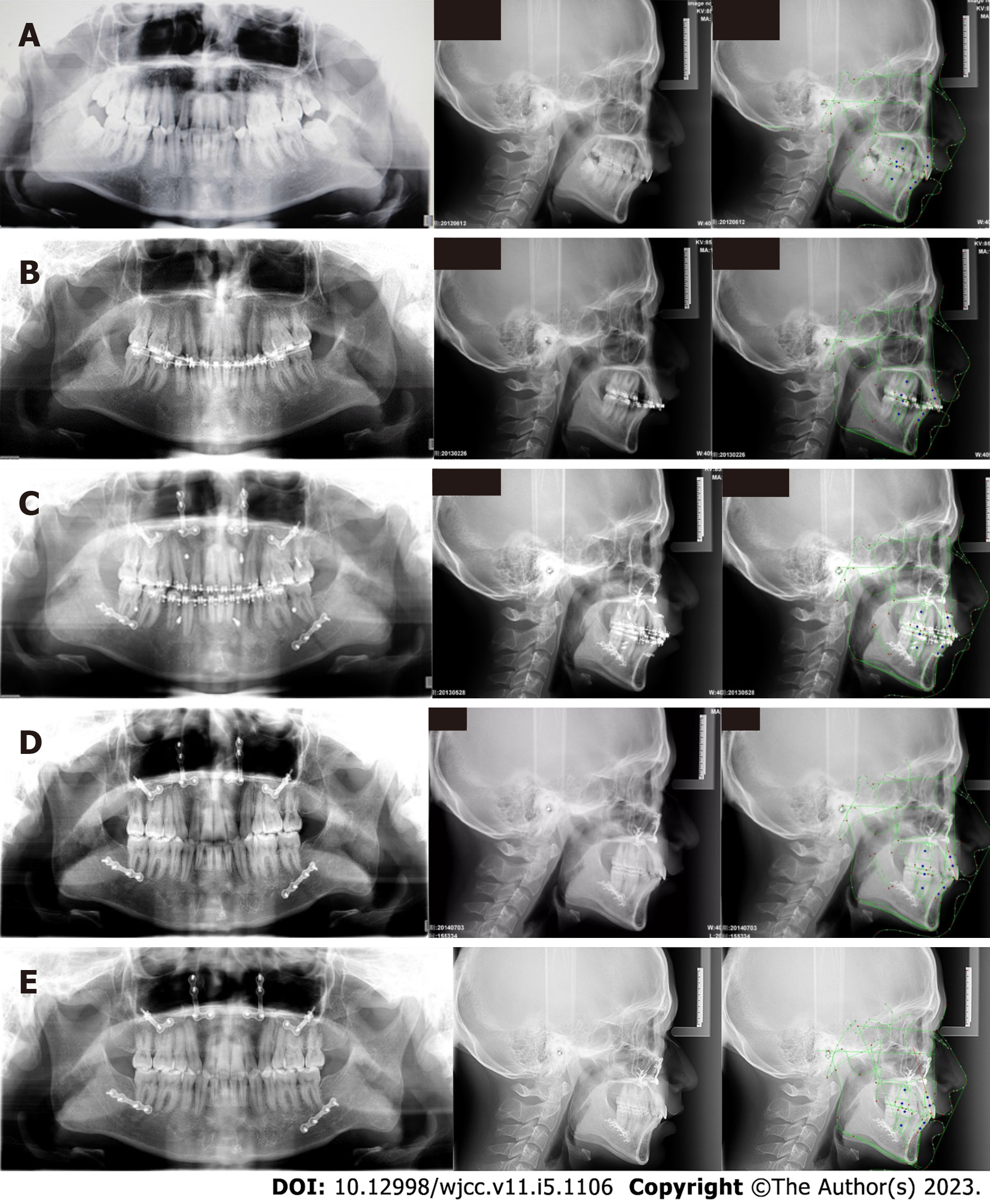
Figure 3 Radiographs of the patient.
A: Pretreatment radiographs; B: Pre-surgical radiographs; C: Post-surgical radiographs; D: Post-treatment radiographs; E: 12 mo follow-up radiographs.
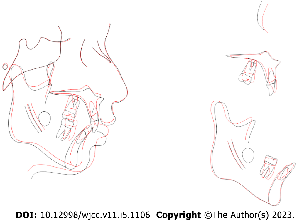
Figure 4 Superimposed tracings.
Black line, pre-treatment; red line, post-treatment.
- Citation: Zhou YW, Wang YY, He ZF, Lu MX, Li GF, Li H. Orthodontic-surgical treatment for severe skeletal class II malocclusion with vertical maxillary excess and four premolars extraction: A case report. World J Clin Cases 2023; 11(5): 1106-1114
- URL: https://www.wjgnet.com/2307-8960/full/v11/i5/1106.htm
- DOI: https://dx.doi.org/10.12998/wjcc.v11.i5.1106


