Copyright
©The Author(s) 2023.
World J Clin Cases. Feb 16, 2023; 11(5): 1086-1093
Published online Feb 16, 2023. doi: 10.12998/wjcc.v11.i5.1086
Published online Feb 16, 2023. doi: 10.12998/wjcc.v11.i5.1086
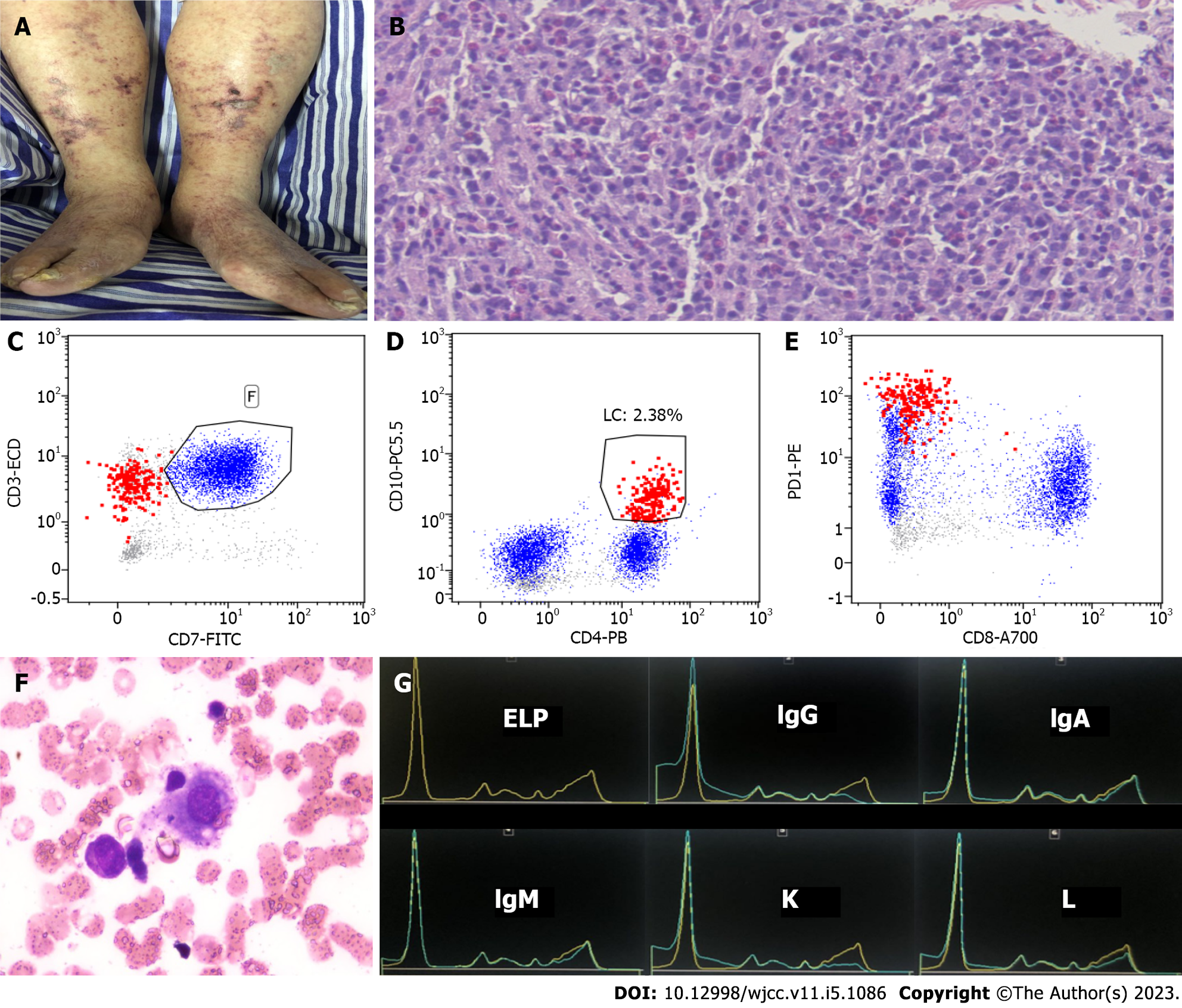
Figure 1 Examinations.
A: Purpura was observed on both lower limbs of the patient; B: Groin lymph node puncture specimen showed that the normal structure of lymph nodes disappeared and heterogeneous infiltration of small to medium-sized lymphoma cells, with proliferation of eosinophils (hematoxylin and eosin staining, × 40); C-E: Flow cytometry. Neoplastic T cells are shown in red and benign T cells in blue (analysis was gating on lymphocytes). The neoplastic T cells were positive for CD3, CD4, CD10, and PD1, but negative for CD7 and CD8; F: Bone marrow examination showed hemophagocytosis; G: Capillary electrophoresis revealed monoclonal IgG kappa.
Figure 2 Positron emission tomography.
A-F: Positron emission tomography showed generalized lymphadenopathy, enhanced activity in the posterior pharyngeal wall (A), bilateral neck (B), hilum of the lung and mediastinum (C), pelvic wall (D), mesenteric lymph nodes (E), and groin (F).
- Citation: Jiang M, Wan JH, Tu Y, Shen Y, Kong FC, Zhang ZL. Angioimmunoblastic T-cell lymphoma induced hemophagocytic lymphohistiocytosis and disseminated intravascular coagulopathy: A case report. World J Clin Cases 2023; 11(5): 1086-1093
- URL: https://www.wjgnet.com/2307-8960/full/v11/i5/1086.htm
- DOI: https://dx.doi.org/10.12998/wjcc.v11.i5.1086


