Copyright
©The Author(s) 2023.
World J Clin Cases. Feb 16, 2023; 11(5): 1031-1039
Published online Feb 16, 2023. doi: 10.12998/wjcc.v11.i5.1031
Published online Feb 16, 2023. doi: 10.12998/wjcc.v11.i5.1031
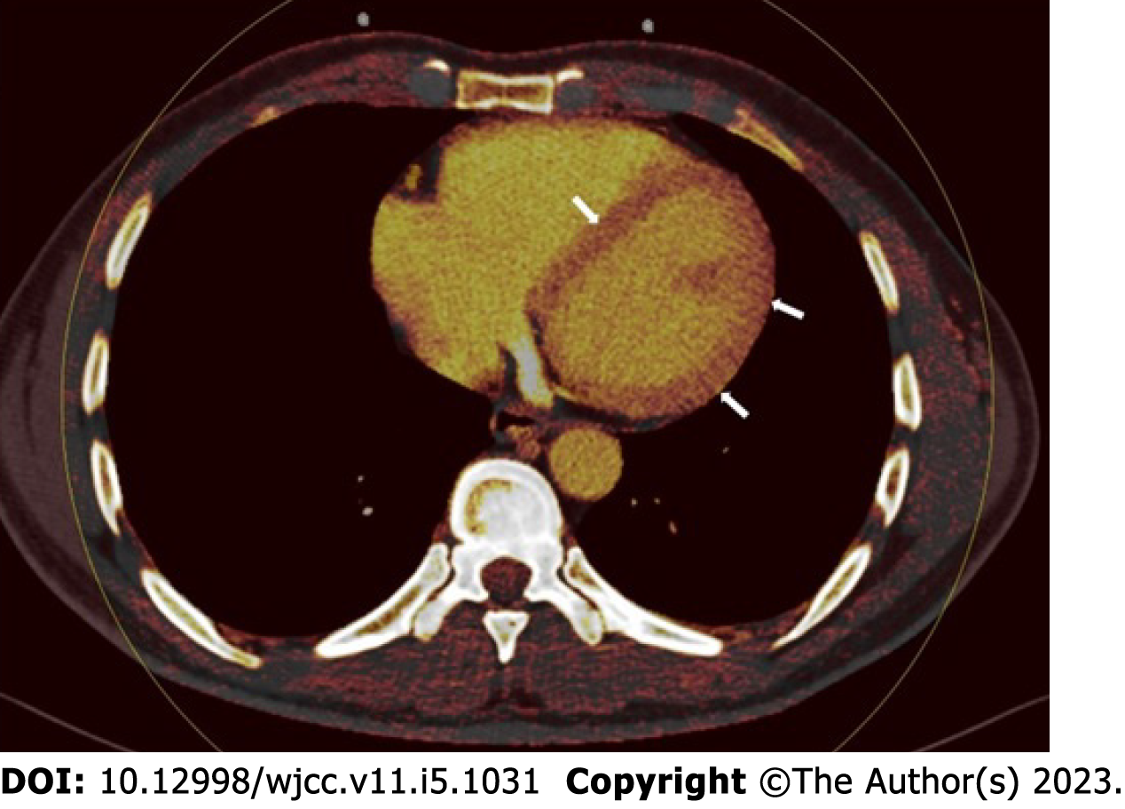
Figure 1 Forty-seven-year-old male with normal left ventricular wall on dual energy computed tomography iodine perfusion map.
Figure 2 Perfusion deficits were found on dual energy computed tomography iodine map images.
A: Fifty-seven-year-old female with corona virus disease 2019 (COVID-19). Subepicardial perfusion deficit at the free wall of the left ventricle on DECT iodine perfusion map (arrows); B: Forty-two-year-old female with COVID-19. Intramyocardial perfusion deficit at the free wall of the left ventricle on dual energy computed tomography (DECT) iodine perfusion map (arrows); C: Fifty-three-year-old male with COVID-19. Transmural perfusion deficit at the free wall of the left ventricle on DECT iodine perfusion map (arrows).
- Citation: Aydin F, Kantarci M, Aydın S, Karavaş E, Ceyhun G, Ogul H, Şahin ÇE, Eren S. COVID-19-related cardiomyopathy: Can dual-energy computed tomography be a diagnostic tool? World J Clin Cases 2023; 11(5): 1031-1039
- URL: https://www.wjgnet.com/2307-8960/full/v11/i5/1031.htm
- DOI: https://dx.doi.org/10.12998/wjcc.v11.i5.1031


