Copyright
©The Author(s) 2023.
World J Clin Cases. Dec 26, 2023; 11(36): 8535-8541
Published online Dec 26, 2023. doi: 10.12998/wjcc.v11.i36.8535
Published online Dec 26, 2023. doi: 10.12998/wjcc.v11.i36.8535
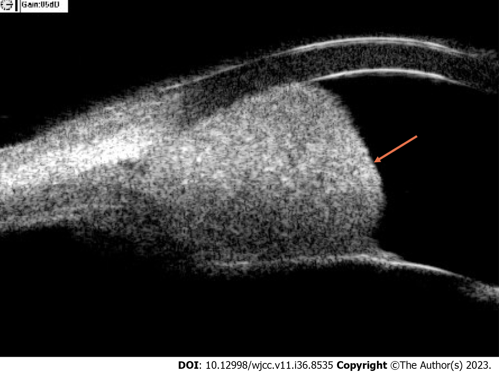
Figure 1 Eye ultrasound image.
A well-bounded, high-density lesion was observed at the corner of the anterior chamber at the 3 o’clock position.
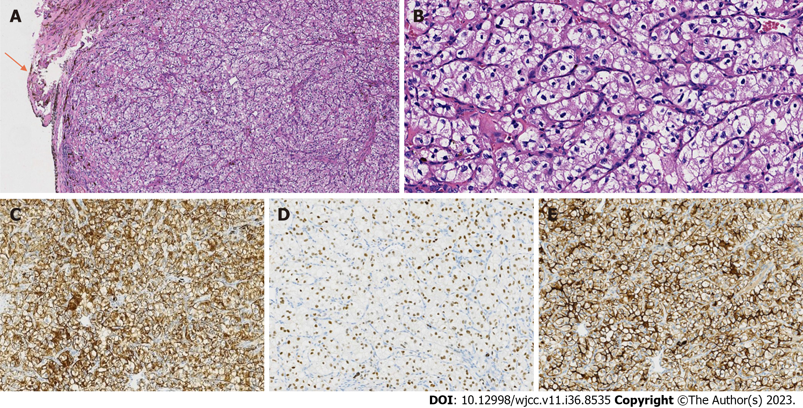
Figure 2 Hematoxylin and eosin staining and immunohistochemical staining of the Iris mass.
A: Normal iris tissue was observed around the tumor (100 ×); B: The tumor cells were large, cube-shaped, and in a solid nest-like arrangement and the nuclei were round or ovate with visible nucleoli (400 ×); C-E: The tumor cells were immunoreactive for cytokeratin pan (200 ×) (C), paired box gene 8 (200 ×) (D), and cluster of differentiation 10 (200 ×) (E).
- Citation: Wang TT, Chen XY, Min QY, Han YZ, Zhao HF. Iris metastasis from clear cell renal cell carcinoma: A case report. World J Clin Cases 2023; 11(36): 8535-8541
- URL: https://www.wjgnet.com/2307-8960/full/v11/i36/8535.htm
- DOI: https://dx.doi.org/10.12998/wjcc.v11.i36.8535


