Copyright
©The Author(s) 2023.
World J Clin Cases. Dec 6, 2023; 11(34): 8170-8175
Published online Dec 6, 2023. doi: 10.12998/wjcc.v11.i34.8170
Published online Dec 6, 2023. doi: 10.12998/wjcc.v11.i34.8170
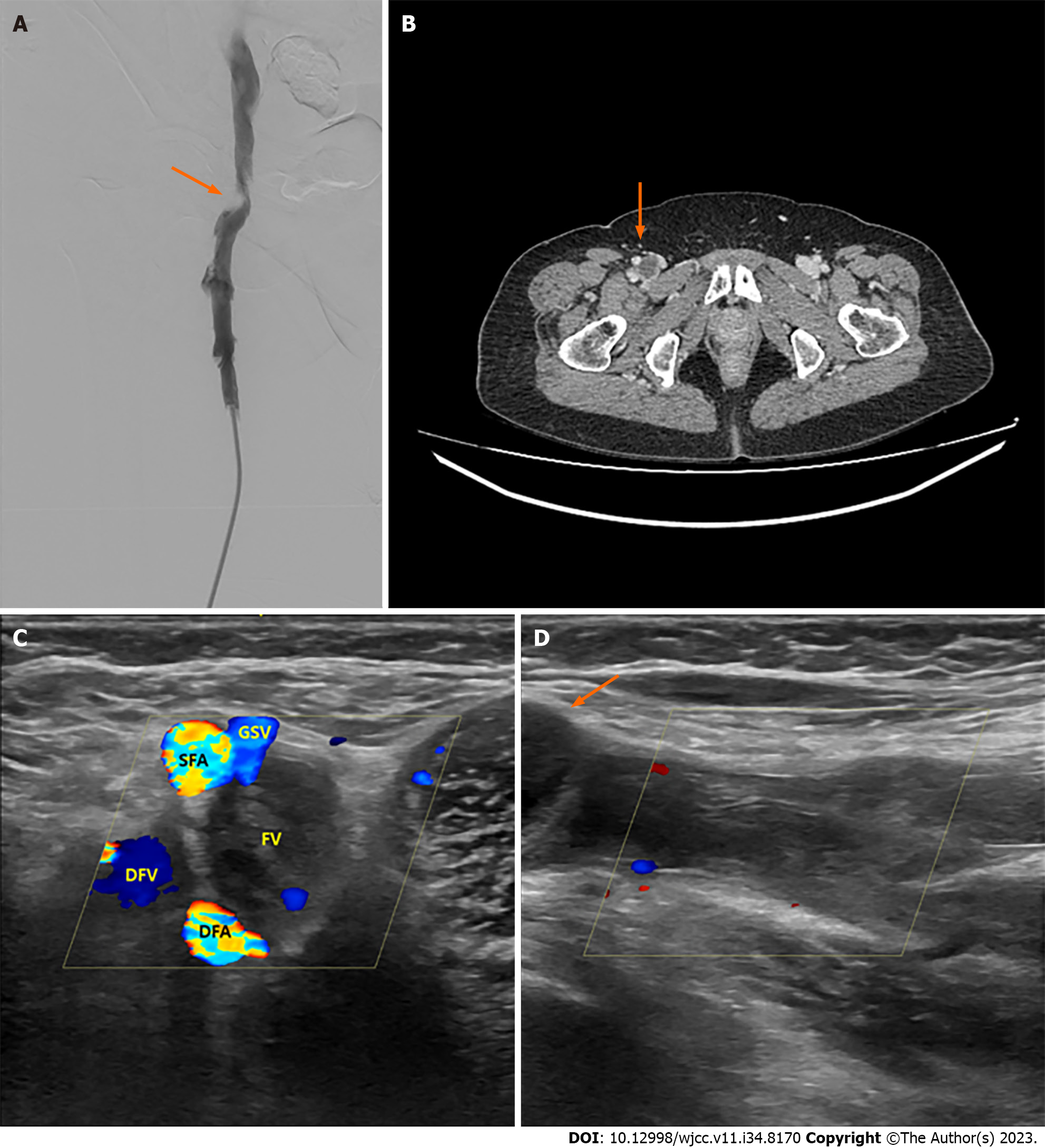
Figure 1 Image study.
A: Venography showing a mass-like lesion (scimitar sign) outside the blood vessel pressing on the right common femoral vein (FV) (CFV) (arrow); B: The cystic mass pressing on the right CFV (arrow); C and D: On ultrasonography, flow to the right FV cannot be observed because of a cystic mass blocking the right CFV; C: Transverse view; D: Longitudinal view. Arrows point to the cyst.
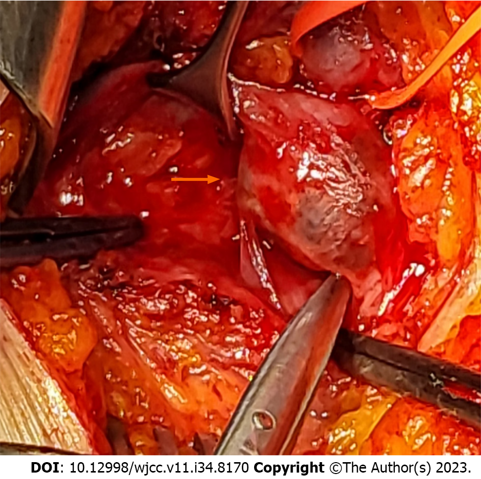
Figure 2 Right common femoral vein.
Arrow: Venous adv.
- Citation: Bae M, Huh U, Lee CW, Kim JW. Venous adventitial cystic disease is a very rare disease that can cause deep vein thrombosis: A case report. World J Clin Cases 2023; 11(34): 8170-8175
- URL: https://www.wjgnet.com/2307-8960/full/v11/i34/8170.htm
- DOI: https://dx.doi.org/10.12998/wjcc.v11.i34.8170


