Copyright
©The Author(s) 2023.
World J Clin Cases. Nov 6, 2023; 11(31): 7656-7662
Published online Nov 6, 2023. doi: 10.12998/wjcc.v11.i31.7656
Published online Nov 6, 2023. doi: 10.12998/wjcc.v11.i31.7656
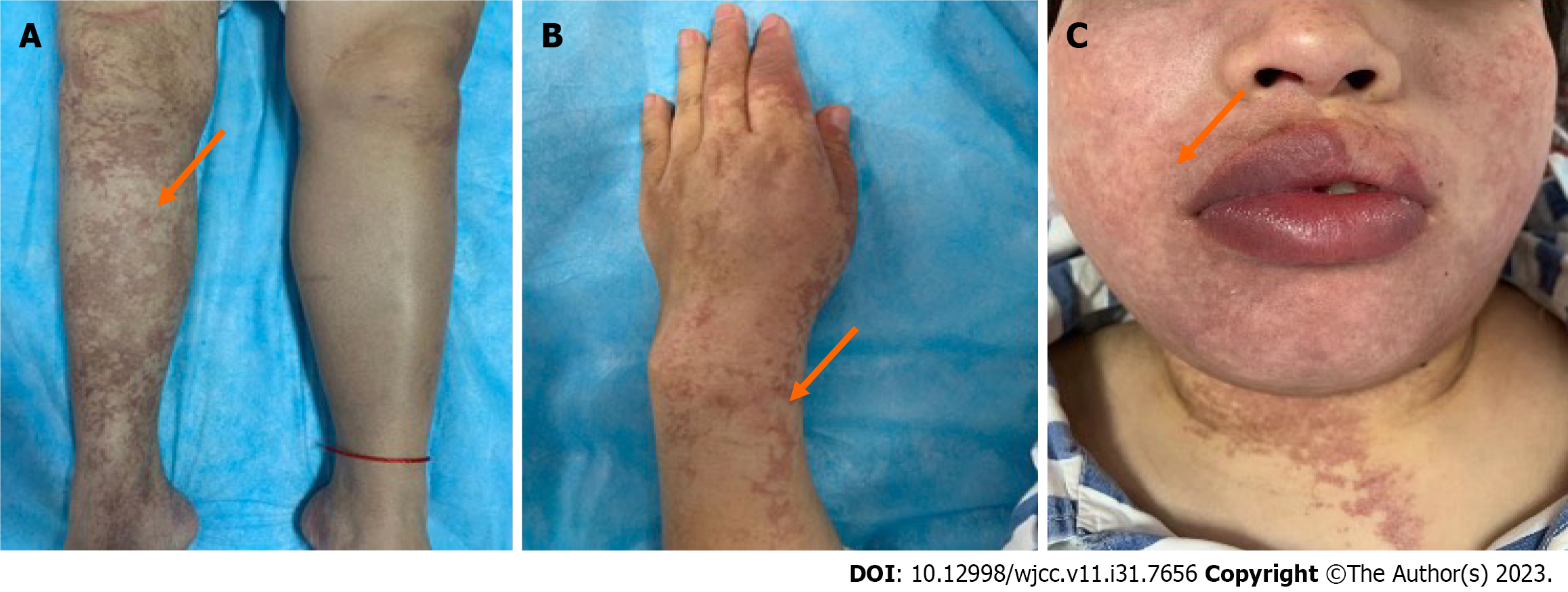
Figure 1 The lesions typically present as irregular, bordered, and ring-shaped bluish-purple patterns.
A: Limbs; B: Hands; C: Face.
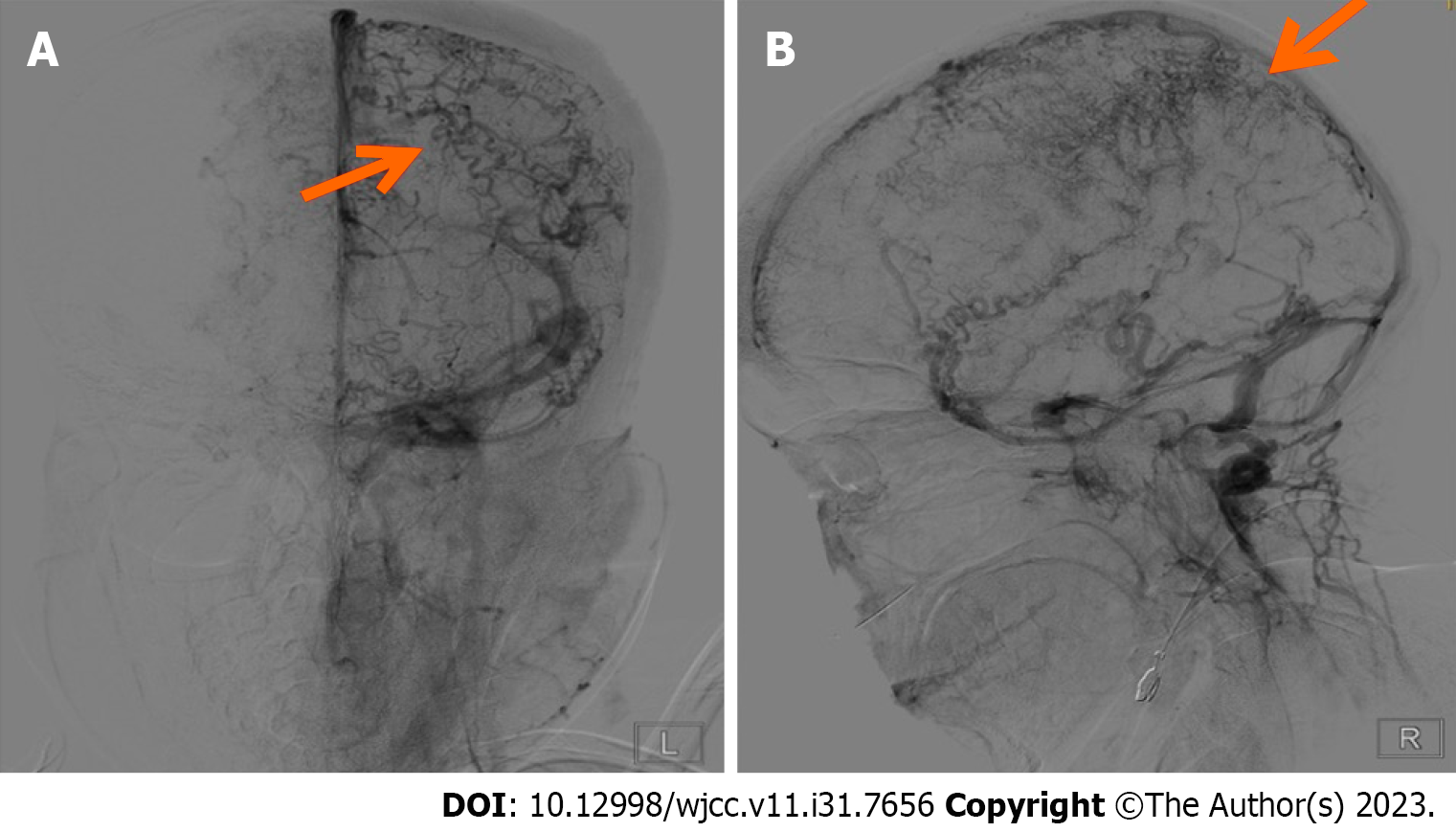
Figure 2 Digital subtraction angiography.
A: The upper sagittal sinuses, lower sagittal sinuses, and straight sinuses were light, and bilateral sigmoid sinuses were tortuous; B: Bilateral cortical veins are extensively tortuous and thickened.
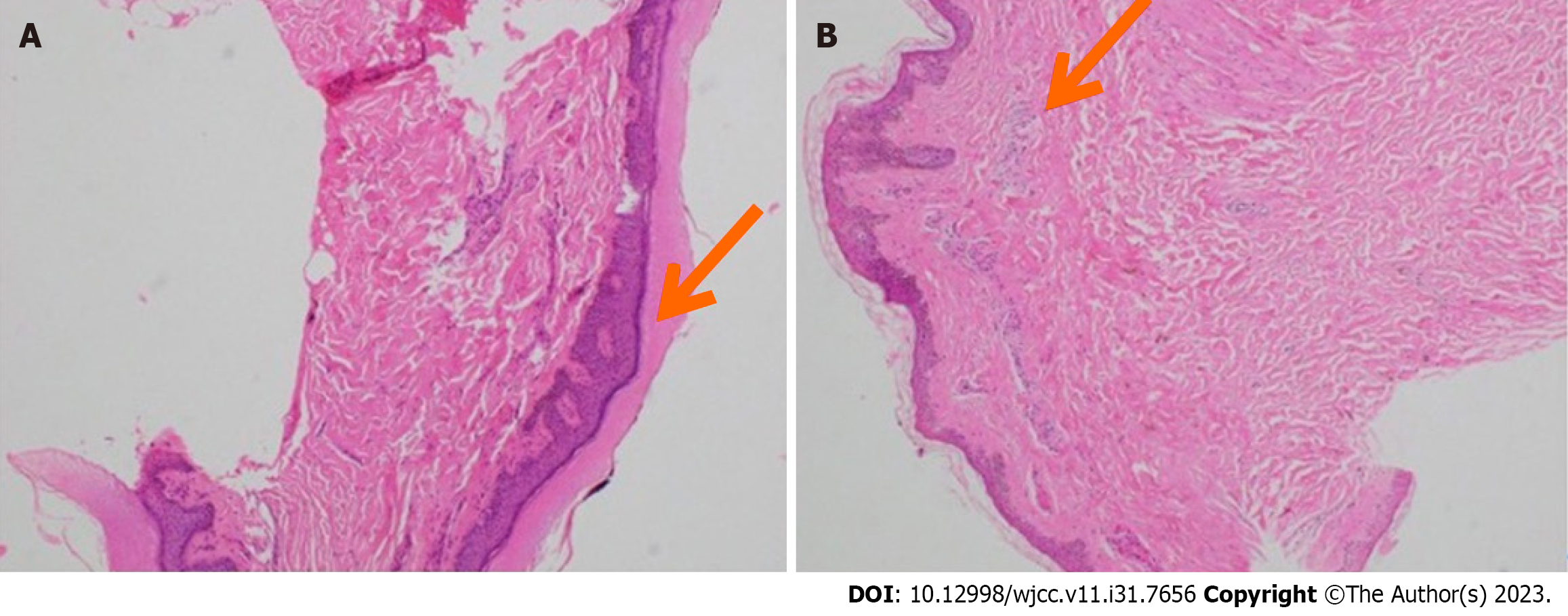
Figure 3 Skin biopsy (hematoxylin-eosin staining).
A: Epidermal hyperkeratosis; B: Subcutaneous chronic inflammatory cell infiltration.
- Citation: Heng Y, Tang YF, Zhang XW, Duan JF, Shi J, Luo Q. Sneddon's syndrome concurrent with cerebral venous sinus thrombosis: A case report. World J Clin Cases 2023; 11(31): 7656-7662
- URL: https://www.wjgnet.com/2307-8960/full/v11/i31/7656.htm
- DOI: https://dx.doi.org/10.12998/wjcc.v11.i31.7656


