Copyright
©The Author(s) 2023.
World J Clin Cases. Oct 26, 2023; 11(30): 7497-7501
Published online Oct 26, 2023. doi: 10.12998/wjcc.v11.i30.7497
Published online Oct 26, 2023. doi: 10.12998/wjcc.v11.i30.7497
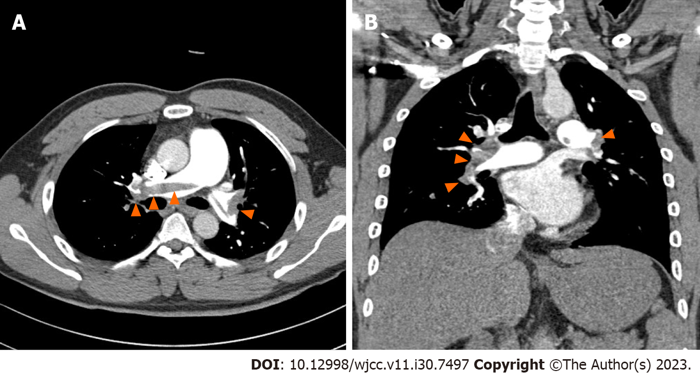
Figure 1 Large amount of contrast-filling defects observed in both the main and lobar segmental pulmonary arteries on contrast-enhanced chest computed tomography.
A: Axial plane; B: Coronal plane.
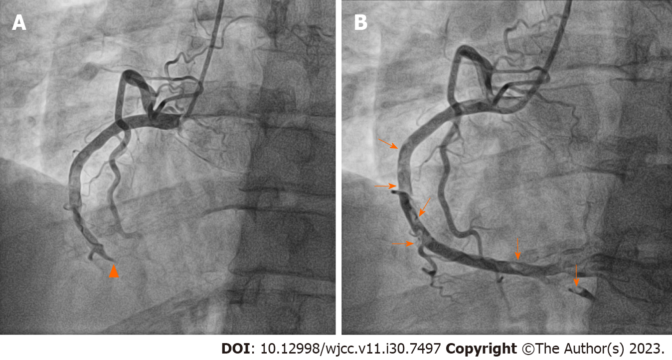
Figure 2 Each figure shows the left-anterior-oblique 30° view on right coronary angiography before and after percutaneous coronary intervention.
A: Coronary angiography revealed a contrast-filling defect lesion in the mid right coronary artery (RCA) without angiographic stenosis (arrowheads) and complete occlusion of the distal RCA; B: Multifocal diffuse thrombi remained in the RCA after percutaneous coronary intervention (arrows).
- Citation: Seo J, Lee J, Shin YH, Jang AY, Suh SY. Acute myocardial infarction after initially diagnosed with unprovoked venous thromboembolism: A case report. World J Clin Cases 2023; 11(30): 7497-7501
- URL: https://www.wjgnet.com/2307-8960/full/v11/i30/7497.htm
- DOI: https://dx.doi.org/10.12998/wjcc.v11.i30.7497


