Copyright
©The Author(s) 2023.
World J Clin Cases. Oct 6, 2023; 11(28): 6902-6907
Published online Oct 6, 2023. doi: 10.12998/wjcc.v11.i28.6902
Published online Oct 6, 2023. doi: 10.12998/wjcc.v11.i28.6902
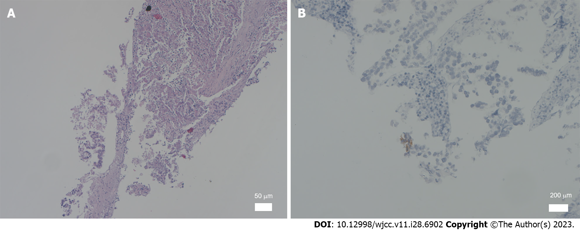
Figure 1 Histological changes and immunohistochemical phenotype of testicular mixed germ cell tumor.
A: Histological changes of malignant germ cell-derived tumor from a male patient (scale bar: 50 μm); B: Immunohistochemical staining for cluster of differentiation 117 (scale bar: 200 μm).
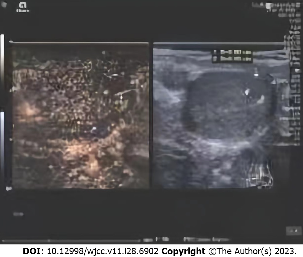
Figure 2 Contrast-enhanced ultrasound findings.
The images show hypoechoic areas in the right testis with poorly defined boundaries and irregular shapes, with short strips of strong echoes.
- Citation: Xiao QF, Li J, Tang B, Zhu YQ. Testicular mixed germ cell tumor: A case report. World J Clin Cases 2023; 11(28): 6902-6907
- URL: https://www.wjgnet.com/2307-8960/full/v11/i28/6902.htm
- DOI: https://dx.doi.org/10.12998/wjcc.v11.i28.6902


