Copyright
©The Author(s) 2023.
World J Clin Cases. Oct 6, 2023; 11(28): 6871-6876
Published online Oct 6, 2023. doi: 10.12998/wjcc.v11.i28.6871
Published online Oct 6, 2023. doi: 10.12998/wjcc.v11.i28.6871
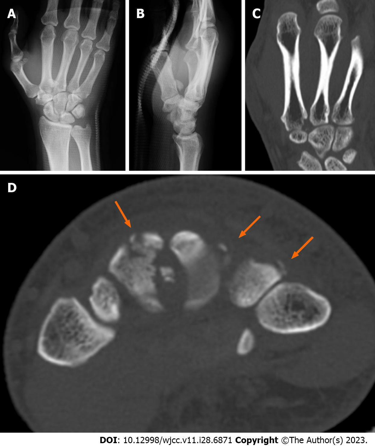
Figure 1 Initial imaging examinations.
A: The initial anteroposterior radiograph shows a displaced fracture fragment between the bases of the second and third metacarpals; B: The initial lateral radiograph shows the fragment lying over the volar aspect of the carpometacarpal joint; C: Coronal view of the initial reformatted multiplanar computed tomography image shows that approximately one-third of the carpometacarpal joint surface of the second metacarpal is damaged; D: Horizontal view of the initial computed tomography image shows small avulsion fragments on the dorsal aspect of the second to fourth metacarpal bases (orange arrows).
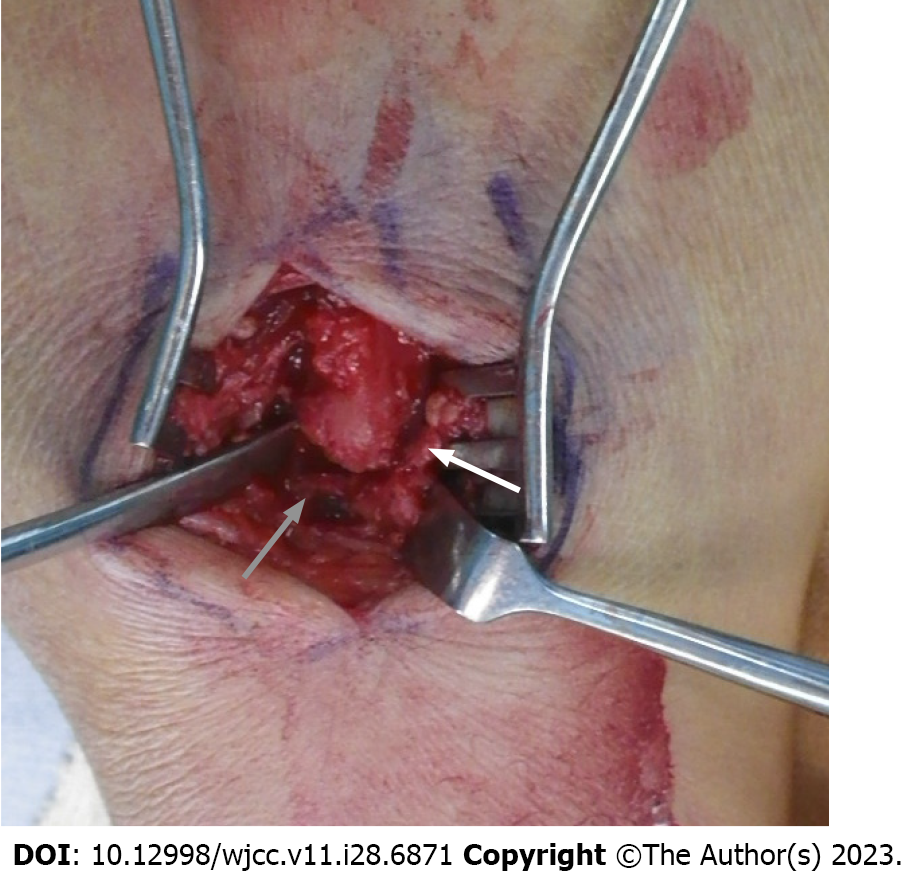
Figure 2
Intraoperative macroscopic photo shows that the dorsal ligament of the third carpometacarpal joint (gray arrow) and the interosseous ligament between the third and fourth metacarpals are torn (white arrow).
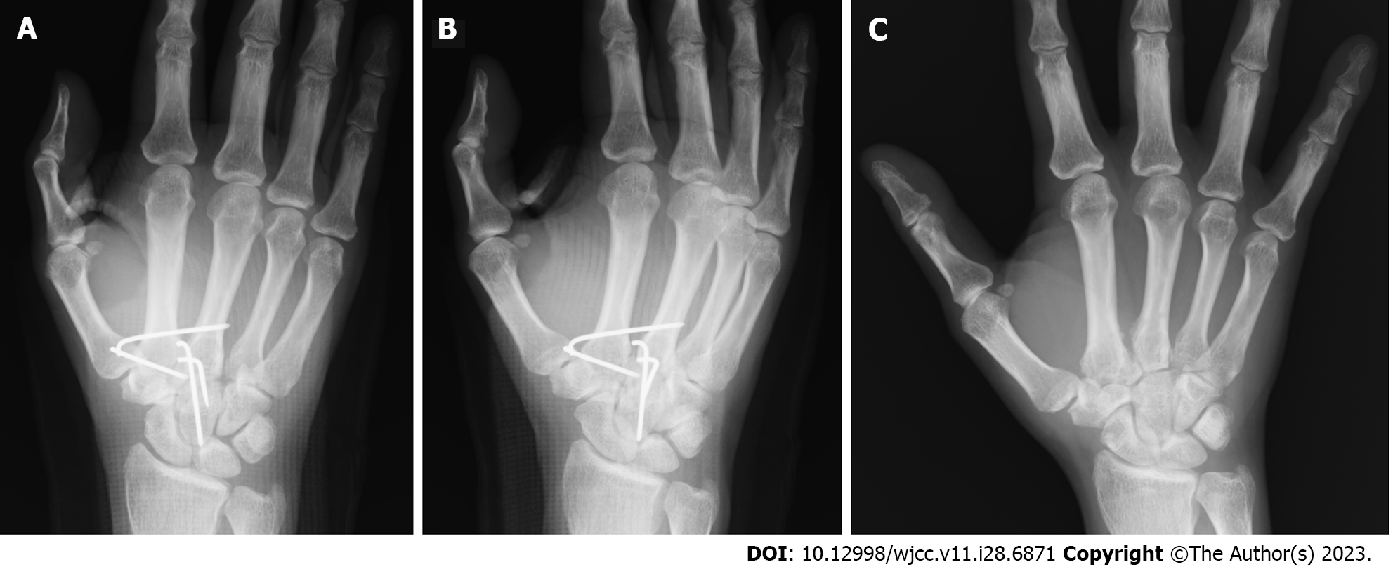
Figure 3 Postoperative radiographs.
A: Anteroposterior postoperative radiograph shows that the fragment is reduced to its anatomical position and stabilized using Kirschner wires; B: Oblique postoperative radiograph shows that the fragment is reduced to its anatomical position and stabilized using Kirschner wires; C: Anteroposterior radiograph at postoperative 12 wk shows union of the second metacarpal fragments.
- Citation: Kurozumi T, Saito M, Odachi K, Masui F. Dorsal approach for isolated volar fracture-dislocation of the base of the second metacarpal: A case report. World J Clin Cases 2023; 11(28): 6871-6876
- URL: https://www.wjgnet.com/2307-8960/full/v11/i28/6871.htm
- DOI: https://dx.doi.org/10.12998/wjcc.v11.i28.6871


