Copyright
©The Author(s) 2023.
World J Clin Cases. Oct 6, 2023; 11(28): 6817-6822
Published online Oct 6, 2023. doi: 10.12998/wjcc.v11.i28.6817
Published online Oct 6, 2023. doi: 10.12998/wjcc.v11.i28.6817
Figure 1 Photos of the child at different ages.
A-C: Photos of the child at the ages of 3 (A), 5 (B), and 8 (C) years.
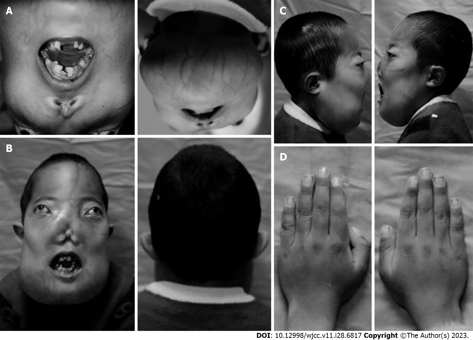
Figure 2 Physical examination images.
A: 45° head up image; B: Front and back images; C: Profile; D: Image of both hands.
Figure 3 Abnormal proliferation of multiple bone fibers throughout the body, including the tibia, femur, humerus, ulna, radius, and skull.
A: X-ray examination of the head; B: X-ray examination of the entire spine; C: X-ray examination of the lower limbs.
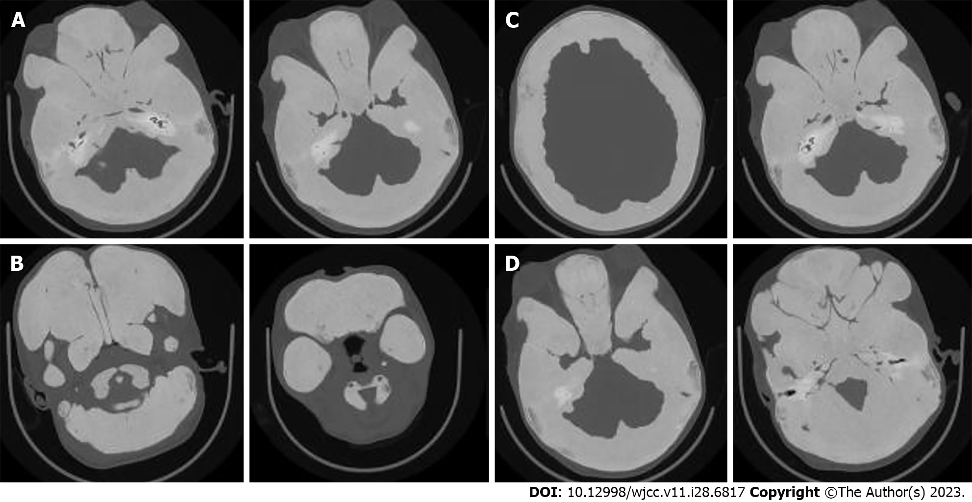
Figure 4 Temporal bone computed tomography examination.
A: Bilateral middle ear with narrow tympanic cavity and sinus; B: No gasification was found in the bilateral maxillary sinuses, ethmoid sinuses, frontal sinuses, sphenoid sinuses, and mastoid chambers; C: Diffuse thickening and increased density of the maxillofacial bone, skull, and atlas axis; D: Narrow bone parts of the external auditory meatus on both sides.
- Citation: Lin X, Feng NY, Lei YJ. Diagnosis and treatment of McCune-Albright syndrome: A case report. World J Clin Cases 2023; 11(28): 6817-6822
- URL: https://www.wjgnet.com/2307-8960/full/v11/i28/6817.htm
- DOI: https://dx.doi.org/10.12998/wjcc.v11.i28.6817


