Copyright
©The Author(s) 2023.
World J Clin Cases. Sep 26, 2023; 11(27): 6664-6669
Published online Sep 26, 2023. doi: 10.12998/wjcc.v11.i27.6664
Published online Sep 26, 2023. doi: 10.12998/wjcc.v11.i27.6664
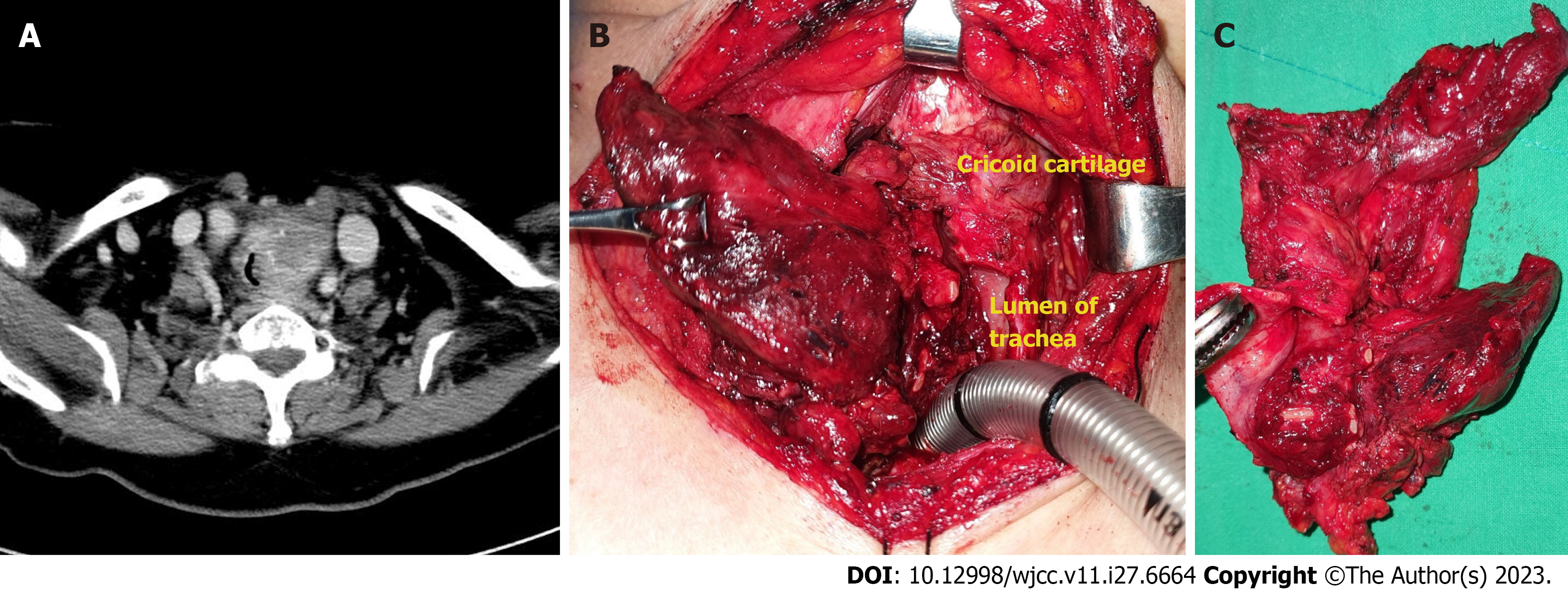
Figure 1 Imaging and findings of the thyroid mass.
A: Neck computed tomography showing a mass of heterogeneous density in the left thyroid gland invading left side of tracheal wall; B and C: Gross invasion of the thyroid mass into the surrounding structures was noted during surgery.
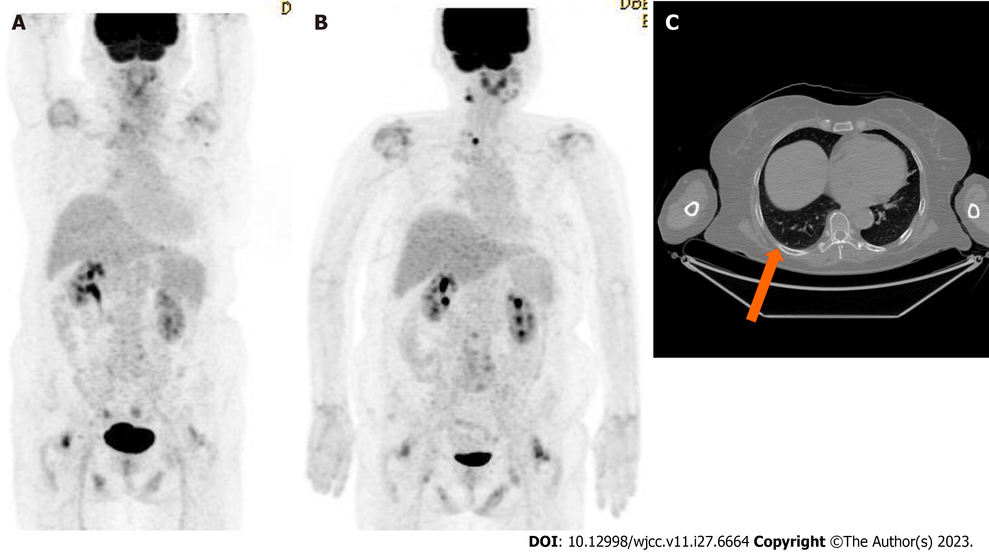
Figure 2 Positron emission tomography/computed tomography findings.
A: Initial positron emission tomography/computed tomography (PET/CT) showing no distant metastasis; B: PET/CT performed at two months post-surgery showing newly developed lesions in the right levels II and VI, along with a pulmonary nodule in the right lower lobe; C: PET/CT showing a 3 mm metastatic pulmonary nodule (orange arrow) in the right lower lobe.
- Citation: Lee SJ, Song SY, Kim MK, Na HG, Bae CH, Kim YD, Choi YS. Complete response of metastatic BRAF V600-mutant anaplastic thyroid cancer following adjuvant dabrafenib and trametinib treatment: A case report. World J Clin Cases 2023; 11(27): 6664-6669
- URL: https://www.wjgnet.com/2307-8960/full/v11/i27/6664.htm
- DOI: https://dx.doi.org/10.12998/wjcc.v11.i27.6664


