Copyright
©The Author(s) 2023.
World J Clin Cases. Sep 26, 2023; 11(27): 6551-6557
Published online Sep 26, 2023. doi: 10.12998/wjcc.v11.i27.6551
Published online Sep 26, 2023. doi: 10.12998/wjcc.v11.i27.6551
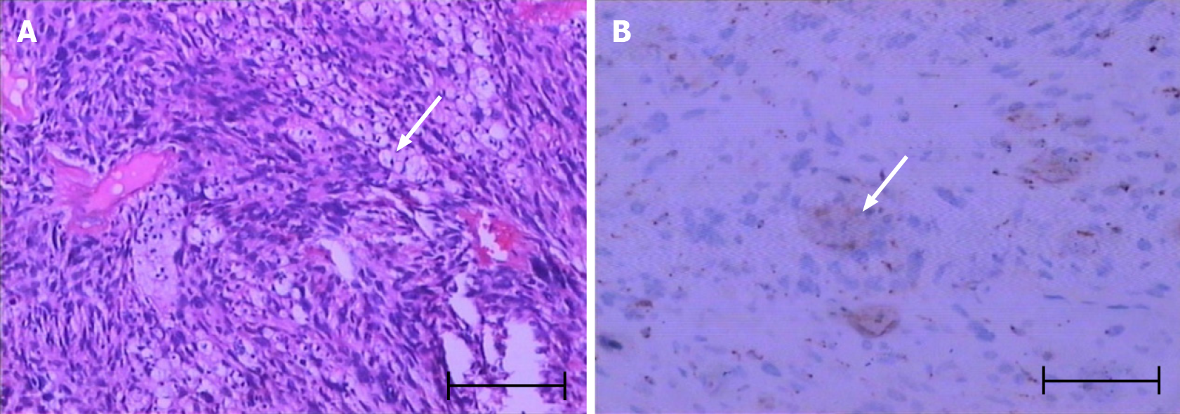
Figure 1 Pathological examination and immunohistochemical detection.
A: H&E staining showing obviously heterogeneous and fat spindle-shaped cells, arranged in intertwined and bundle-like patterns (arrow); B: Immunohistochemical staining establishing the spindle cell origin of the abnormal cell population: CD68+ (arrow). Scale bar: 100 μm.
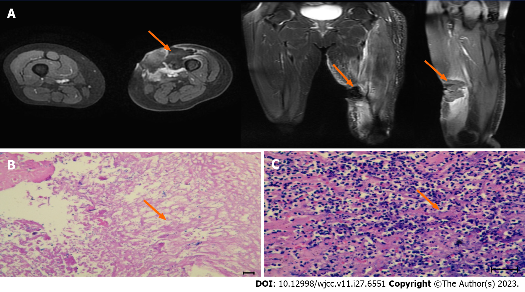
Figure 2 Magnetic resonance imaging imaging and pathological biopsy after the first high-intensity focused ultrasound treatment.
A: An ovoid mass (about 26 mm × 36 mm × 36 mm) with an equal/slightly low signal on T1WI and a slightly low/high mixed signal on T2WI was seen under the skin of the anterior medial part of the left mid-thigh, with a clear border. The lesion was mildly enhanced at the edge after enhancement. A patchy slightly high signal on T2WI with poorly defined borders was seen adjacent to the left middle femur (about 11 mm × 14 mm × 55 mm), with heterogeneous enhancement after enhancement; B and C: H&E staining showing acute and chronic inflammation with necrotic granulomatous tissue proliferation. Scale bar: 100 μm.
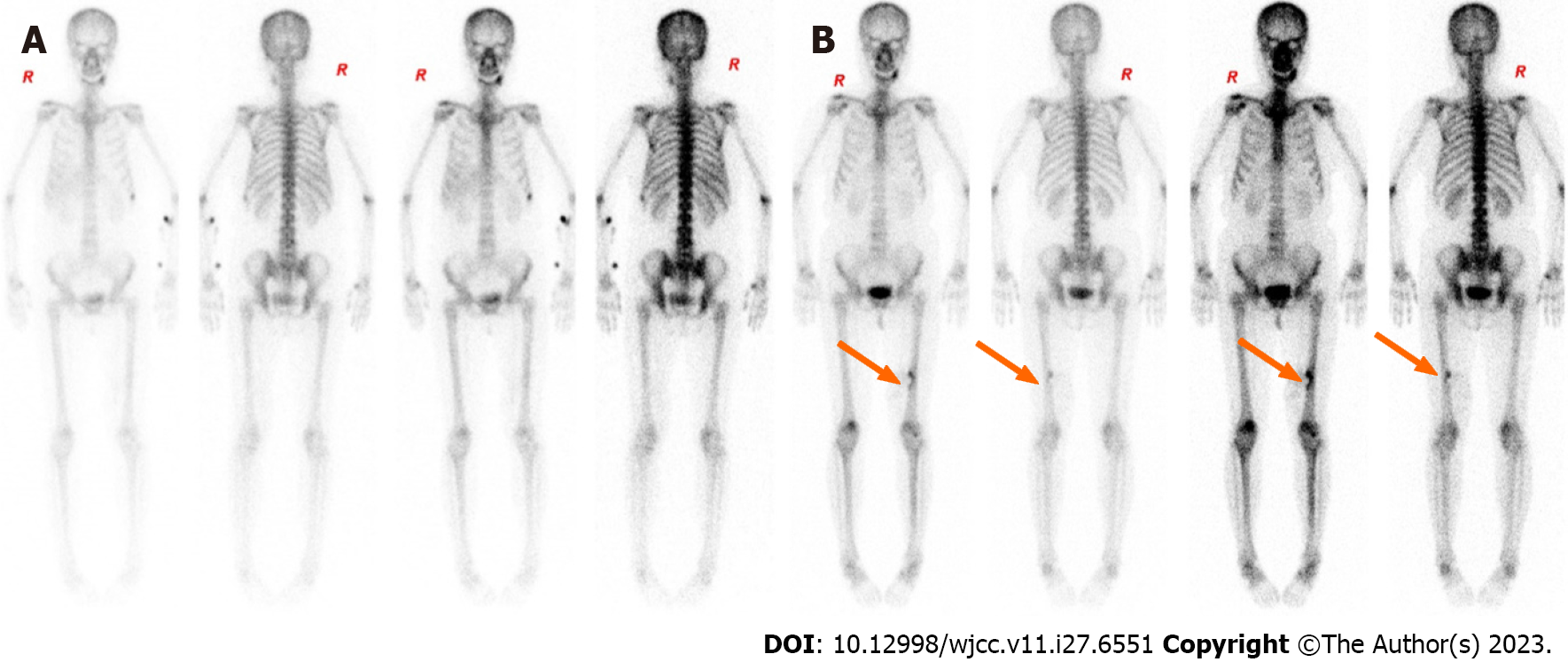
Figure 3 Comparison whole-body scintigraphy before and after high-intensity focused ultrasound treatment.
A: Before high-intensity focused ultrasound (HIFU) (October 19, 2020): No abnormalities; B: After HIFU (June 7, 2021): Localized radiolucent defect at the inner edge of the left lower and middle femoral segments involving a marginal increase in radiolucent shadow, suggesting post-HIFU changes (arrows).
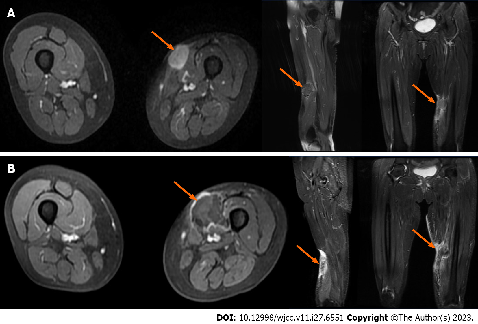
Figure 4 Comparison of magnetic resonance imaging of lower limbs before and after high-intensity focused ultrasound treatment.
A and B: Enhanced magnetic resonance imaging (MRI) of the left thigh. A: Before high-intensity focused ultrasound (HIFU) (March 1, 2021): On the coronal, sagittal and cross-sectional MRI of the left thigh, an oval signal nodule with slightly longer T1 and slightly longer T2 (about 31 mm × 26 mm × 30 mm) was found under the skin of the anterior medial side of the middle part of the left thigh (arrows). After enhancement, it was obviously slightly uneven, and the surrounding soft tissues were edema; B: After HIFU (March 12, 2021): On the coronal, sagittal and cross-sectional MRI of the left thigh, the oval signal nodule with slightly longer T1 and slightly longer T2 (about 28 mm × 30 mm × 25 mm) is located under the skin of the anterior medial side of the middle part of the left thigh, and its boundary with subcutaneous fat and adjacent muscle tissue is unclear (arrows).
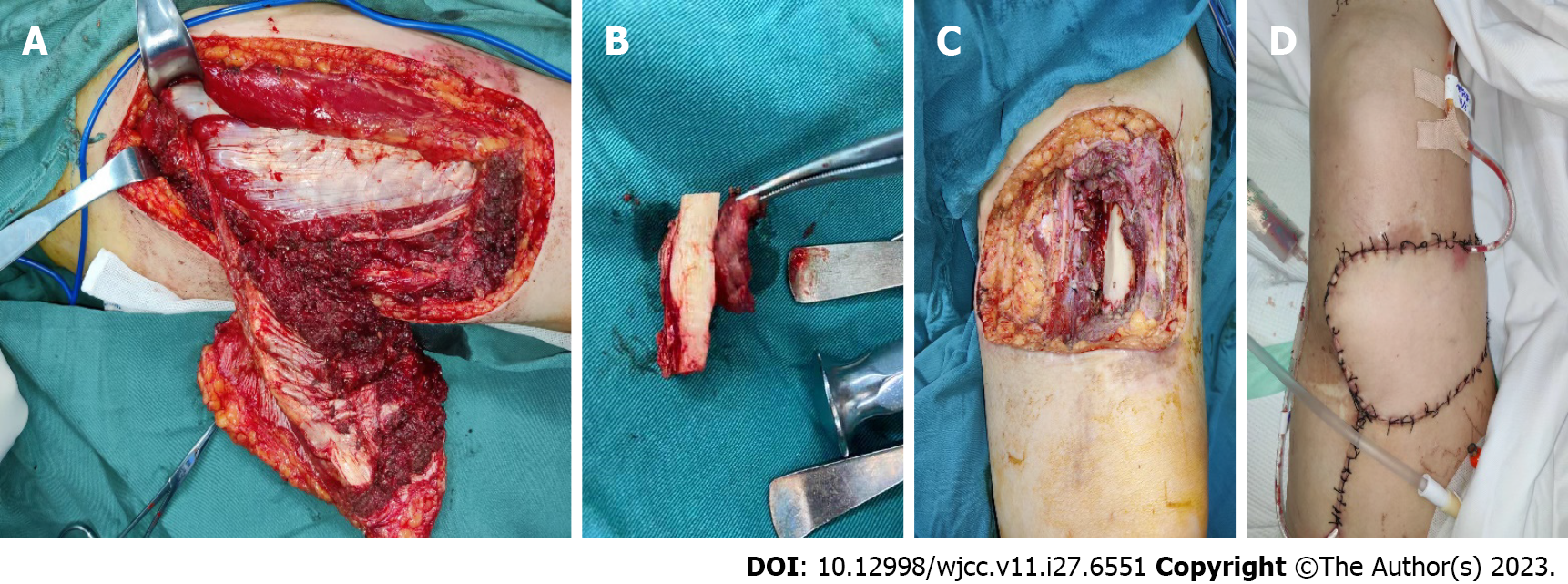
Figure 5 Illustration of patient undergoing surgery on March 3, 2022.
A–D: Surgical images. A: A tissue breaks down (about 8 cm × 10 cm × 5 cm) was seen above the knee joint on the left thigh, deep to the medial, lateral, and rectus femoris muscles; B: Trauma after excision of the mass; C: Left thigh swelling: Gray/yellow/brown soft tissue with skin (10 cm × 6 cm × 3.5 cm) and a gray/white/brown mass (4 cm × 3.5 cm × 2.5 cm) on the skin surface; D: The wound was closed with an 8 cm × 6 cm flap and a drainage tube was left in place.
- Citation: Zhu YQ, Zhao GC, Zheng CX, Yuan L, Yuan GB. Managing spindle cell sarcoma with surgery and high-intensity focused ultrasound: A case report. World J Clin Cases 2023; 11(27): 6551-6557
- URL: https://www.wjgnet.com/2307-8960/full/v11/i27/6551.htm
- DOI: https://dx.doi.org/10.12998/wjcc.v11.i27.6551


