Copyright
©The Author(s) 2023.
World J Clin Cases. Sep 16, 2023; 11(26): 6289-6297
Published online Sep 16, 2023. doi: 10.12998/wjcc.v11.i26.6289
Published online Sep 16, 2023. doi: 10.12998/wjcc.v11.i26.6289
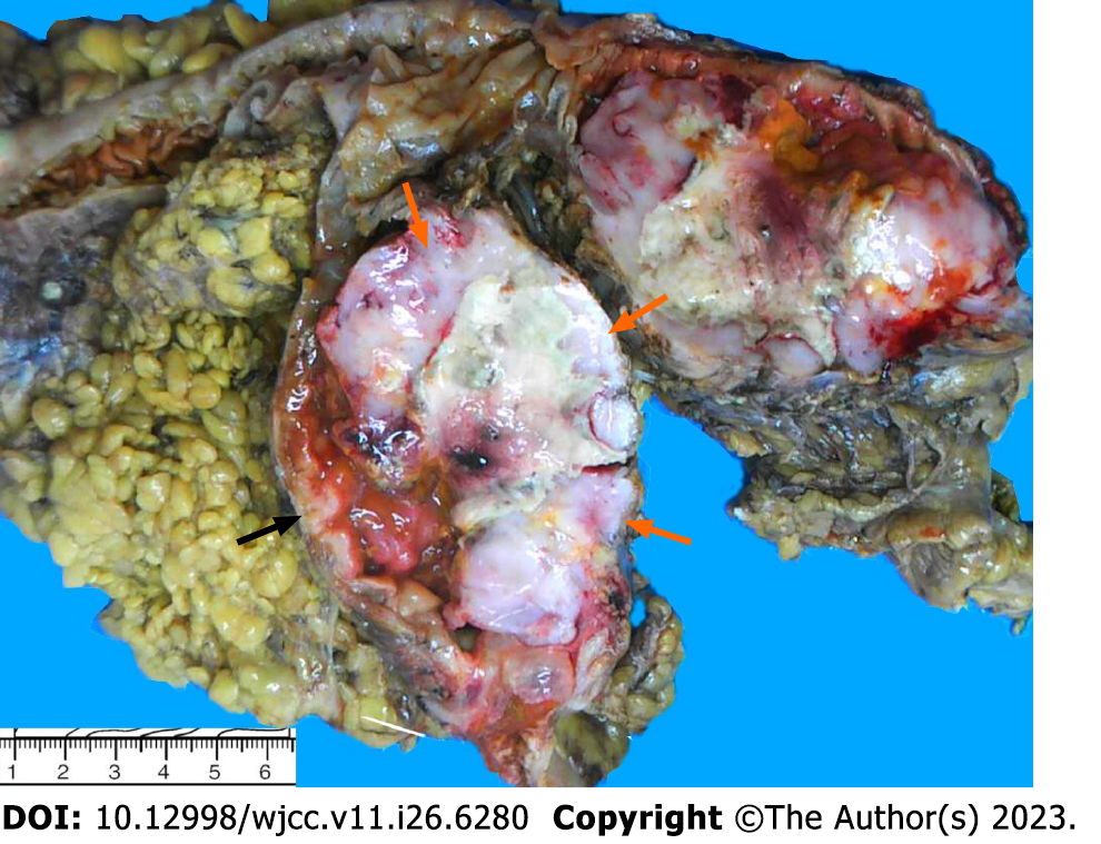
Figure 1 Macroscopic examination.
The resected specimen presented as a circumferential ulcerative mass on the cecal mucosa adjacent to the ileocecal valve. The mass appeared to be comprised of two tumors. The upper-right portion of the mass had a crater-like appearance with a necrotic-appearing central area (orange arrow), whereas the remaining part of the mass had a polypoid, hard and grayish-white aspect (black arrow).
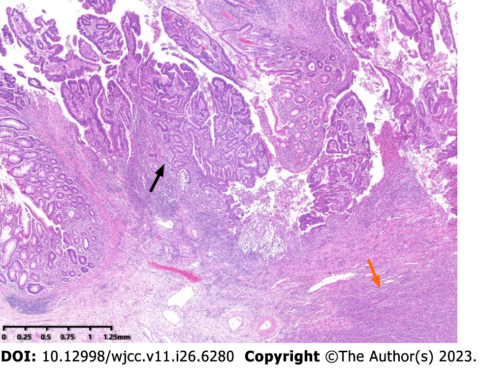
Figure 2 Low power view of the mass.
Microscopic examination disclosed that the tumor was composed of two components, adjacent to each other but relatively independent and showing infiltrative glands (black arrow) with underlying lymphoid proliferation (orange arrow) (Hematoxylin and eosin, original magnificent 20×).
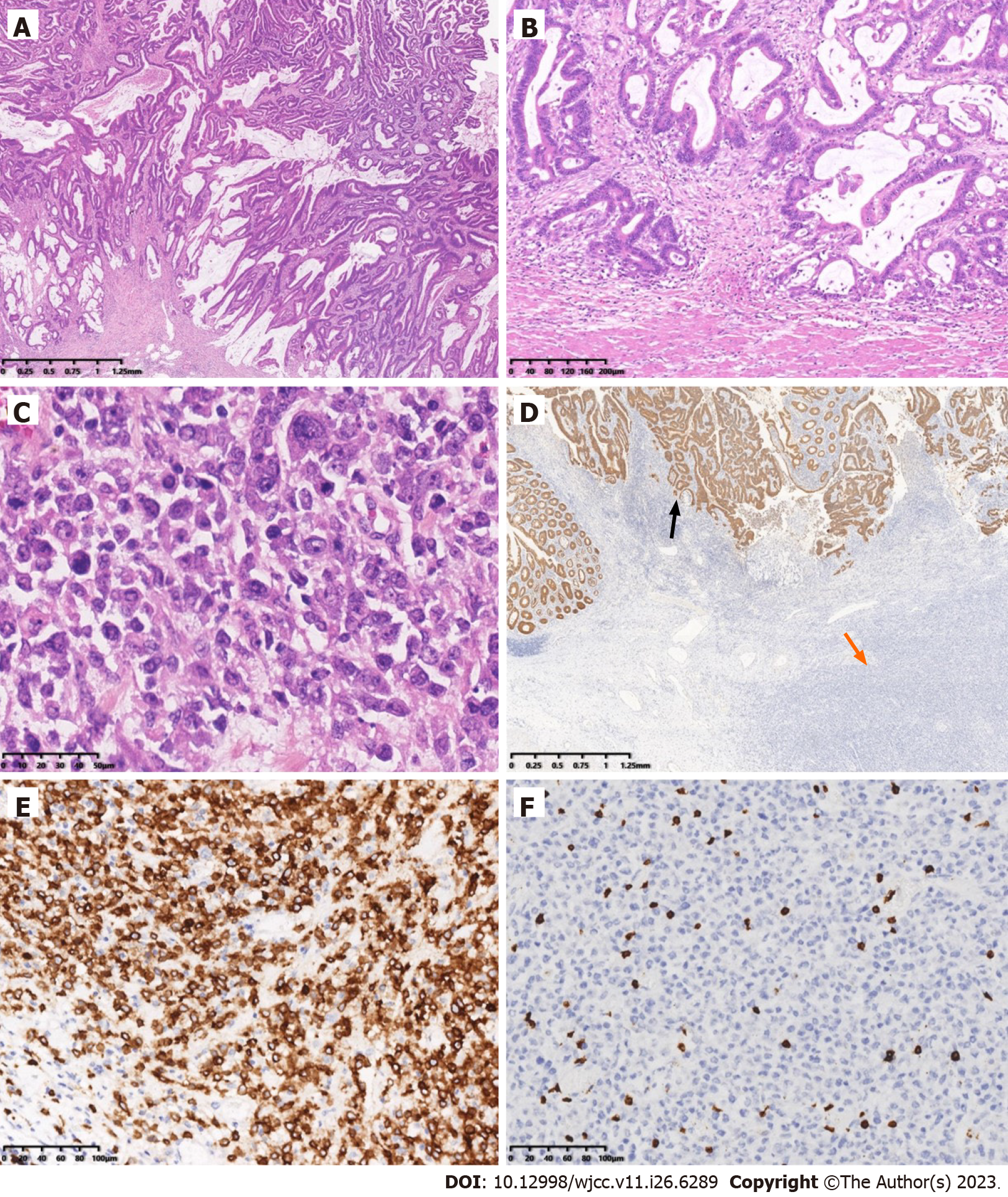
Figure 3 Microscopic examination and immunohistochemistry.
A: Histopathology of one component of the mass was a moderately differentiated adenocarcinoma having a mucinous component arising from a tubulovillous adenoma (top) (Hematoxylin and eosin (HE), original magnification 20×); B: Adenocarcinoma invaded the muscularis propria (HE, original magnification 100×); C: The other component was diffusely medium-to-large lymphocytes, with uniform morphology, frequent mitosis and obvious cell atypia (HE, original magnification 400×); D: Immunohistochemistry of adenocarcinoma was strongly positive for cytokeratin (black arrow), while the atypical lymphocytes were negative (orange arrow) (EnVision method, original magnification 20×); E: Immunohistochemistry of the atypical lymphocytes was strongly and diffusely positive for CD20 (EnVision method, original magnification 200×); F: Immunohistochemistry of the atypical lymphocytes was negative for CD3 (EnVision method, original magnification 200×).
- Citation: Jiang M, Yuan XP. Collision tumor of primary malignant lymphoma and adenocarcinoma in the colon diagnosed by molecular pathology: A case report and literature review. World J Clin Cases 2023; 11(26): 6289-6297
- URL: https://www.wjgnet.com/2307-8960/full/v11/i26/6289.htm
- DOI: https://dx.doi.org/10.12998/wjcc.v11.i26.6289


