Copyright
©The Author(s) 2023.
World J Clin Cases. Sep 6, 2023; 11(25): 5857-5862
Published online Sep 6, 2023. doi: 10.12998/wjcc.v11.i25.5857
Published online Sep 6, 2023. doi: 10.12998/wjcc.v11.i25.5857
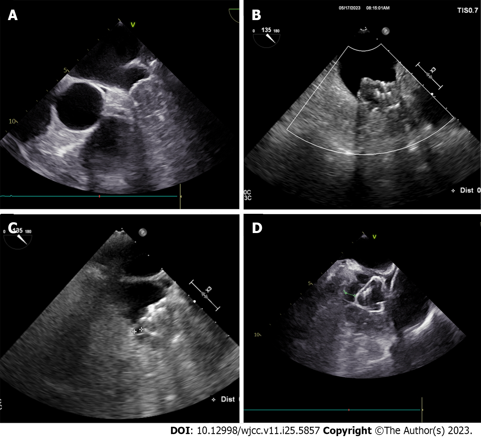
Figure 1 Different grades of peri-device leak using transesophageal echocardiographic imaging.
A: No peri-device leak (PDL); B: 0-3 mm PDL; C: 3-5 mm PDL; D: > 5 mm PDL.
- Citation: Qi YB, Chu HM. Progress in the study and treatment of peri-device leak after left atrial appendage closure. World J Clin Cases 2023; 11(25): 5857-5862
- URL: https://www.wjgnet.com/2307-8960/full/v11/i25/5857.htm
- DOI: https://dx.doi.org/10.12998/wjcc.v11.i25.5857


