Copyright
©The Author(s) 2023.
World J Clin Cases. Jul 6, 2023; 11(19): 4640-4647
Published online Jul 6, 2023. doi: 10.12998/wjcc.v11.i19.4640
Published online Jul 6, 2023. doi: 10.12998/wjcc.v11.i19.4640
Figure 1 Magnetic resonance imaging showed a 36 mm × 28 mm mass under the capsule of the left inner lobe of the liver.
A: No abnormal signal was found in the parenchyma of the remaining liver; B: Retroperitoneal lymphadenopathy was not found; C: Slightly elevated signal intensity on T2-weighted imaging; D: No signs of effusion were found in the abdominal cavity.
Figure 2 The resected part of the liver measured 16 cm × 9 cm × 5 cm.
A: An irregular nodule measuring 3.5 cm × 3.5 cm × 2.5 cm was seen near the resection margin. This nodule was hard, yellow-grey, and well-defined. The tumour did not rupture, and it did not penetrate the liver capsule. The rest of the liver was soft, grey-red, and free of nodules; B: The tumour cells were large, with prominent nucleoli and eosinophilic cytoplasm (H&E, ×200); C: The tumour had an irregular island-like and nest-like morphology, with numerous lymphocytes and plasma cells infiltrating the stroma (H&E, ×200); D: The tumour was infiltrative and indistinguishable from the surrounding liver tissue (H&E, ×200).
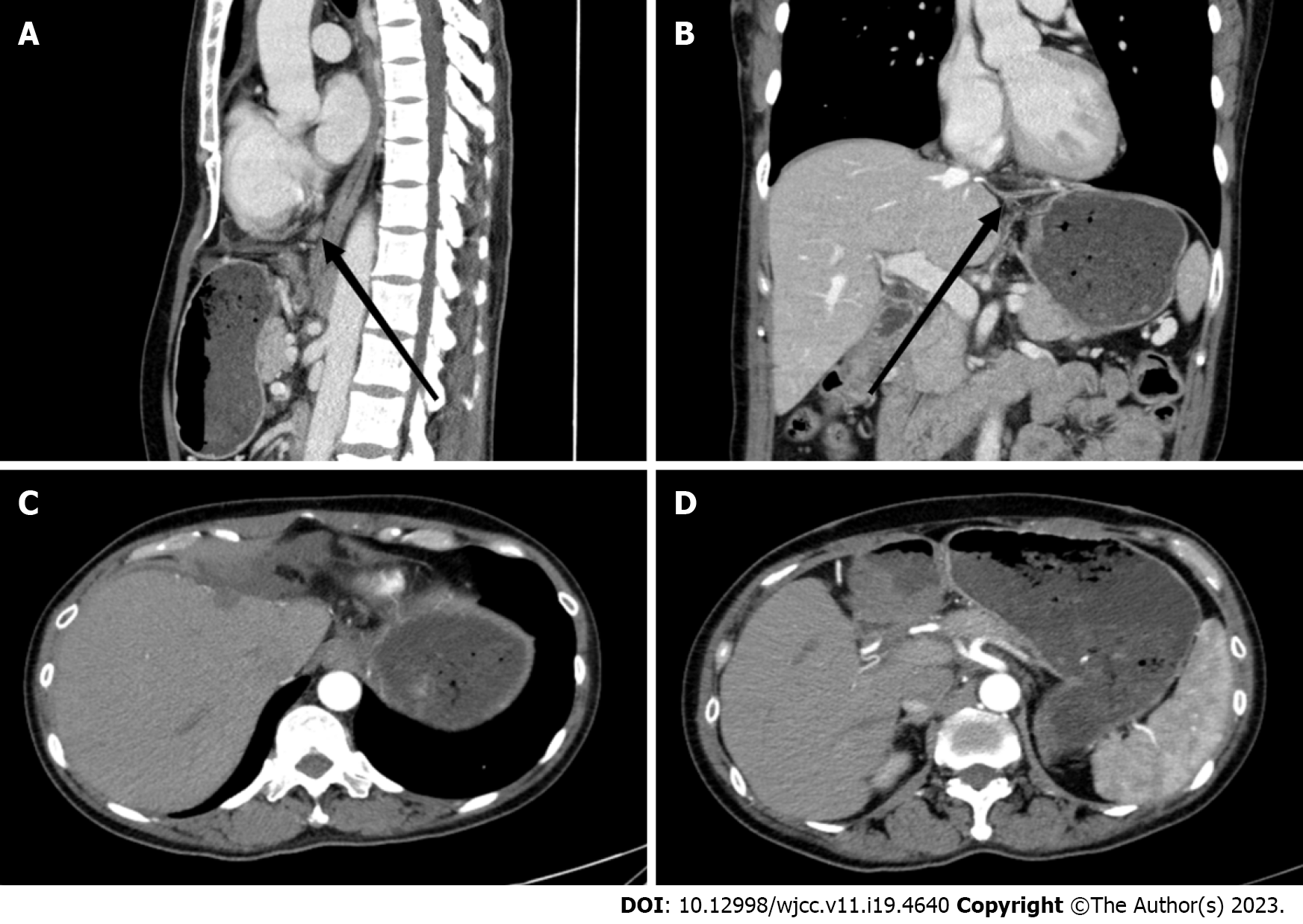
Figure 3 Images of upper abdominal spiral computed tomography performed on December 30, 2022.
A: There were no obvious abnormal lymph nodes in the abdominal cavity or retroperitoneum; B: The left lobe of the liver was absent postoperatively; C: A small amount of hydrops was present in the adjacent abdominal cavity, and there was no obvious widening of the portal vein; D: There was no obvious mass lesion in the operative area.
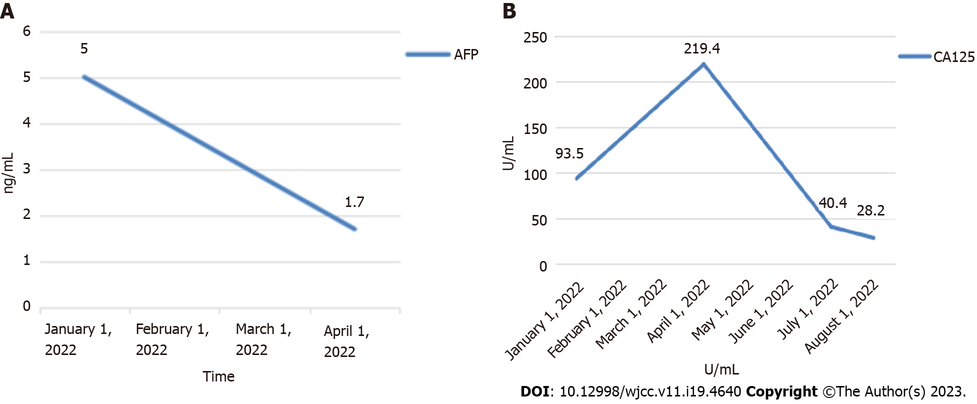
Figure 4 Tumour marker levels.
A: Alpha-fetoprotein decreaseds from 5 ng/mL to 1.7 ng/mL from January 1 to April 1, 2022; B: From January 1 to August 1, 2022, the carbohydrate antigen 125 value decreased from 93.5 U/mL to 28.2 U/mL.
- Citation: Tang HT, Lin W, Zhang WQ, Qian JL, Li K, He K. CK5/6-positive, P63-positive lymphoepithelioma-like hepatocellular carcinoma: A case report and literature review. World J Clin Cases 2023; 11(19): 4640-4647
- URL: https://www.wjgnet.com/2307-8960/full/v11/i19/4640.htm
- DOI: https://dx.doi.org/10.12998/wjcc.v11.i19.4640


