Copyright
©The Author(s) 2023.
World J Clin Cases. Jun 26, 2023; 11(18): 4318-4325
Published online Jun 26, 2023. doi: 10.12998/wjcc.v11.i18.4318
Published online Jun 26, 2023. doi: 10.12998/wjcc.v11.i18.4318
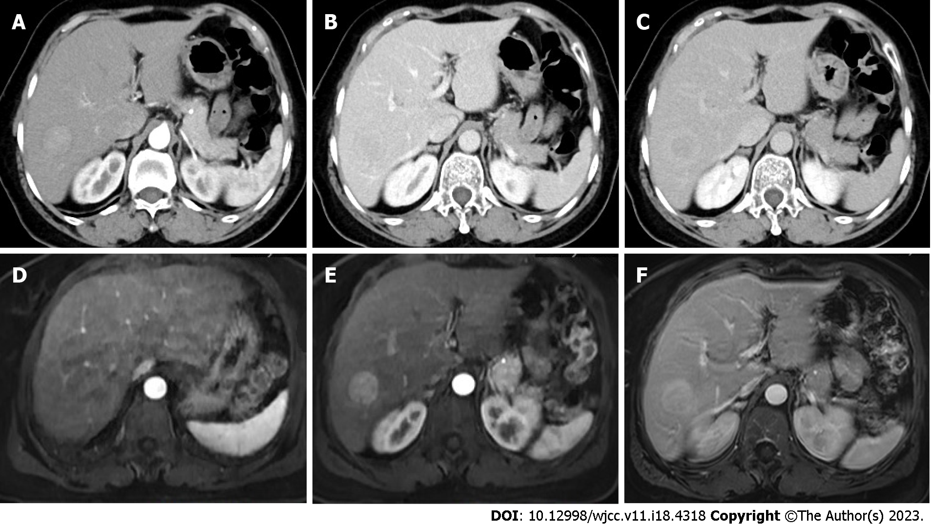
Figure 1 Computed tomography and magnetic resonance imaging revealed a right lower posterior lobe vascular enhancement mass that was suspected to be a malignant tumor.
A-C: Arterial phase, venous phase and delayed phase of enhanced computed tomography showed a fast-in and fast-out typical manifestation of hepatocellular carcinoma; D: Magnetic resonance imaging showed many hepatic nodules of cirrhosis; E and F: Enhanced magnetic resonance imaging showed a typical manifestation of a malignant tumor.
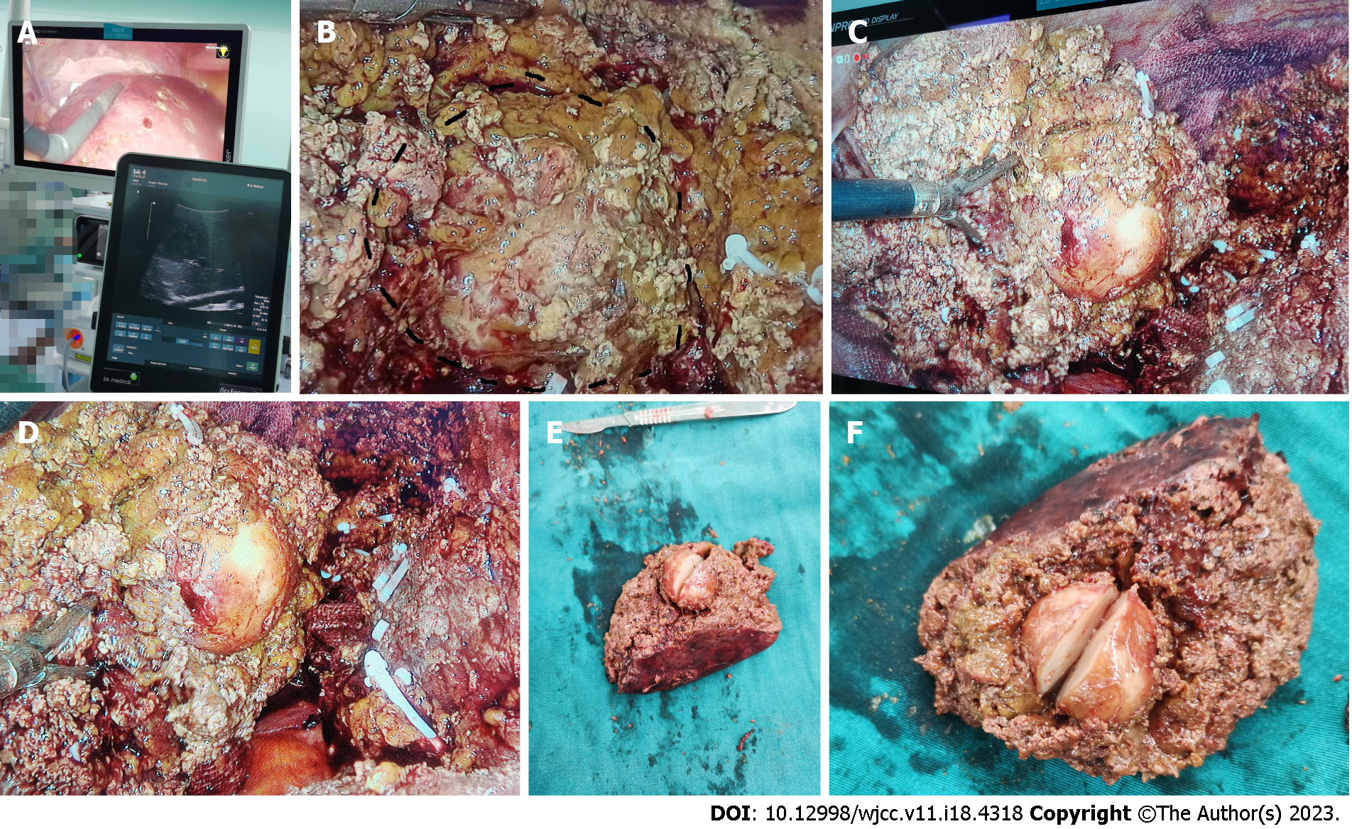
Figure 2 Intraoperative and macroscopic view of liver neoplasm.
A: Ultrasound localization of the edge of the hepatectomy; B-D: Process of liver hepatectomy (the black circle in B shows the site of tumor); E and F: Gross view of the resected liver specimen.
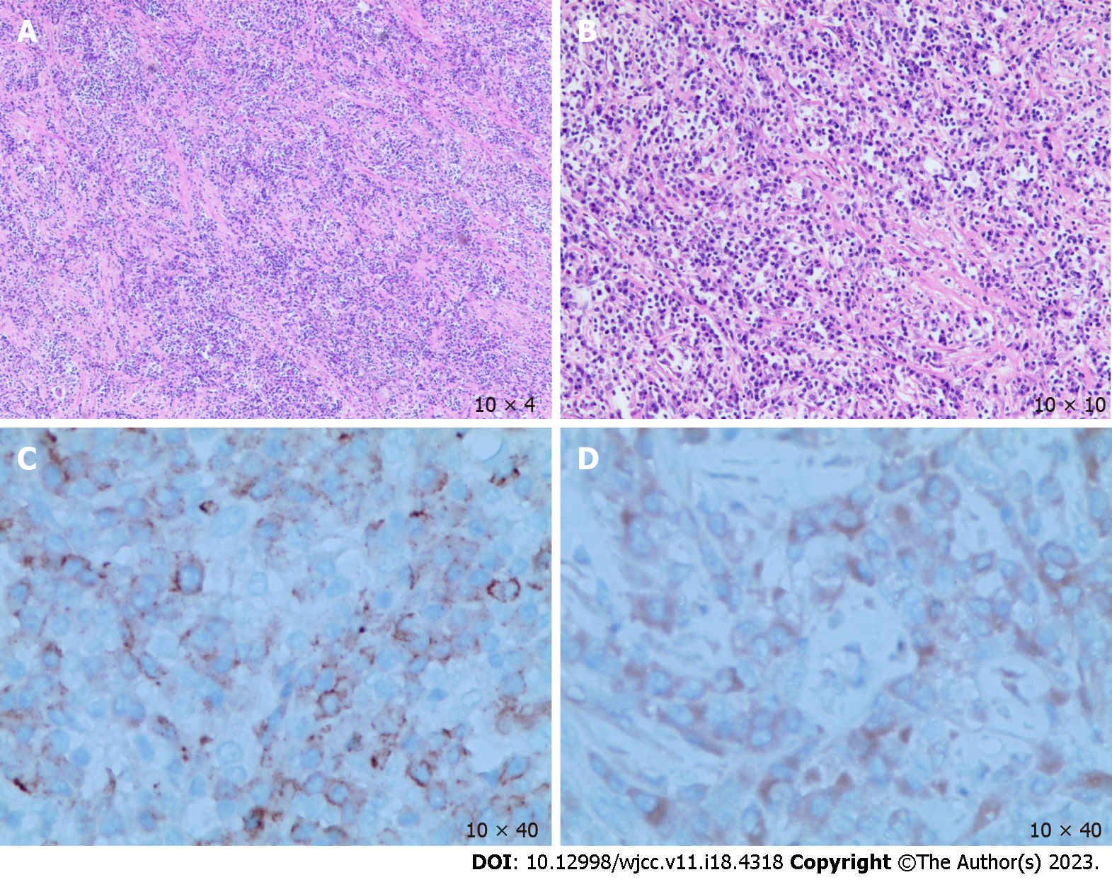
Figure 3 Staining of the specimen.
A and B: Hematoxylin-eosin stain; C: Immunohistochemistry (IHC) stain of CD138; D: IHC stain of anaplastic lymphoma kinase.
- Citation: Tong M, Zhang BC, Jia FY, Wang J, Liu JH. Hepatic inflammatory myofibroblastic tumor: A case report. World J Clin Cases 2023; 11(18): 4318-4325
- URL: https://www.wjgnet.com/2307-8960/full/v11/i18/4318.htm
- DOI: https://dx.doi.org/10.12998/wjcc.v11.i18.4318


