Copyright
©The Author(s) 2023.
World J Clin Cases. Jun 16, 2023; 11(17): 4168-4178
Published online Jun 16, 2023. doi: 10.12998/wjcc.v11.i17.4168
Published online Jun 16, 2023. doi: 10.12998/wjcc.v11.i17.4168
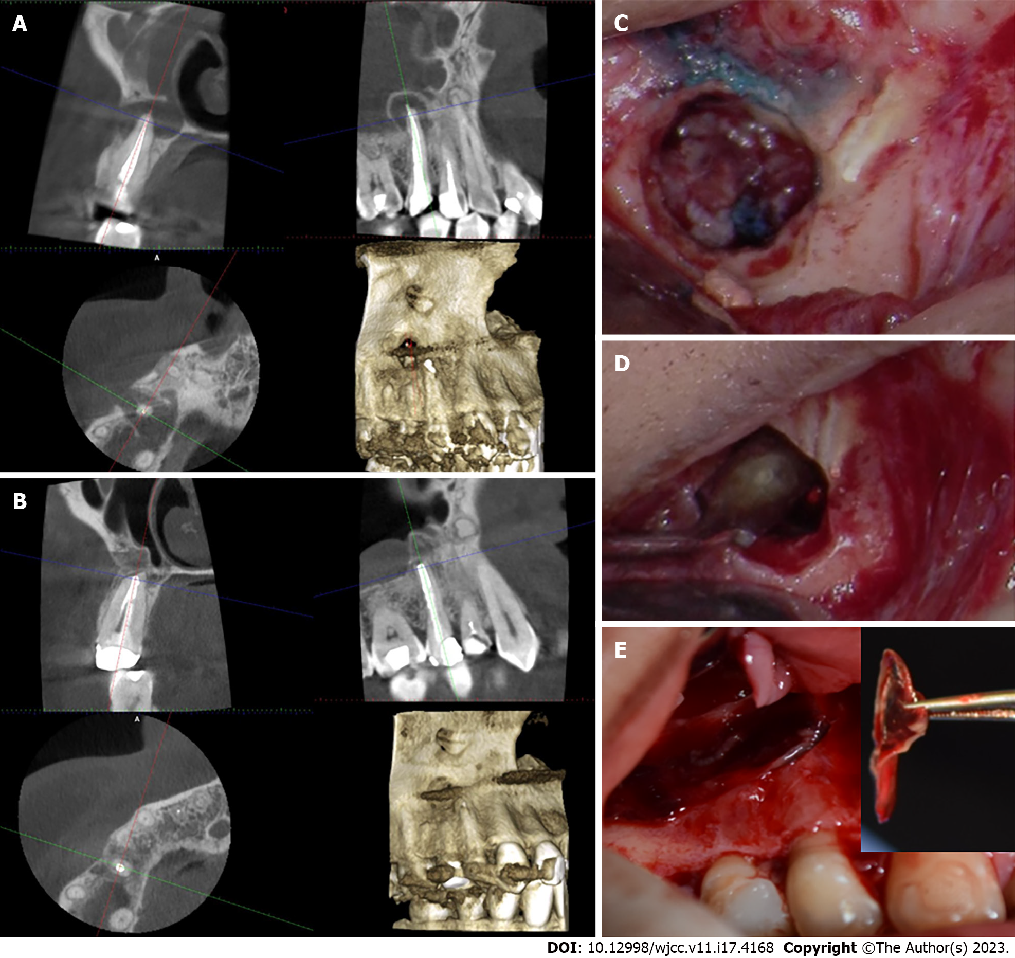
Figure 1 Bone regeneration using advanced platelet-rich fibrin membrane in through-and-through bony defects following periapical endodontic surgery on the maxillary right second premolar.
A: The cone-beam computed tomography (CBCT) shows localized radiolucency surrounding the apex of maxillary right second premolar. The lesion is perforating the labial cortical bone and displaced sinus periosteum upward which is a typical feature of periapical osteoperiostitis. The root canal treatment appears to be overextended with a uniform density; B: The CBCT shows bony healing of the resected area within four months; C: A clinical photo of the resected root; D: A clinical photo of the MTA retro-filling; E: The platelet-rich fibrin membrane handled using a tweezer and placed to seal the bony defect.
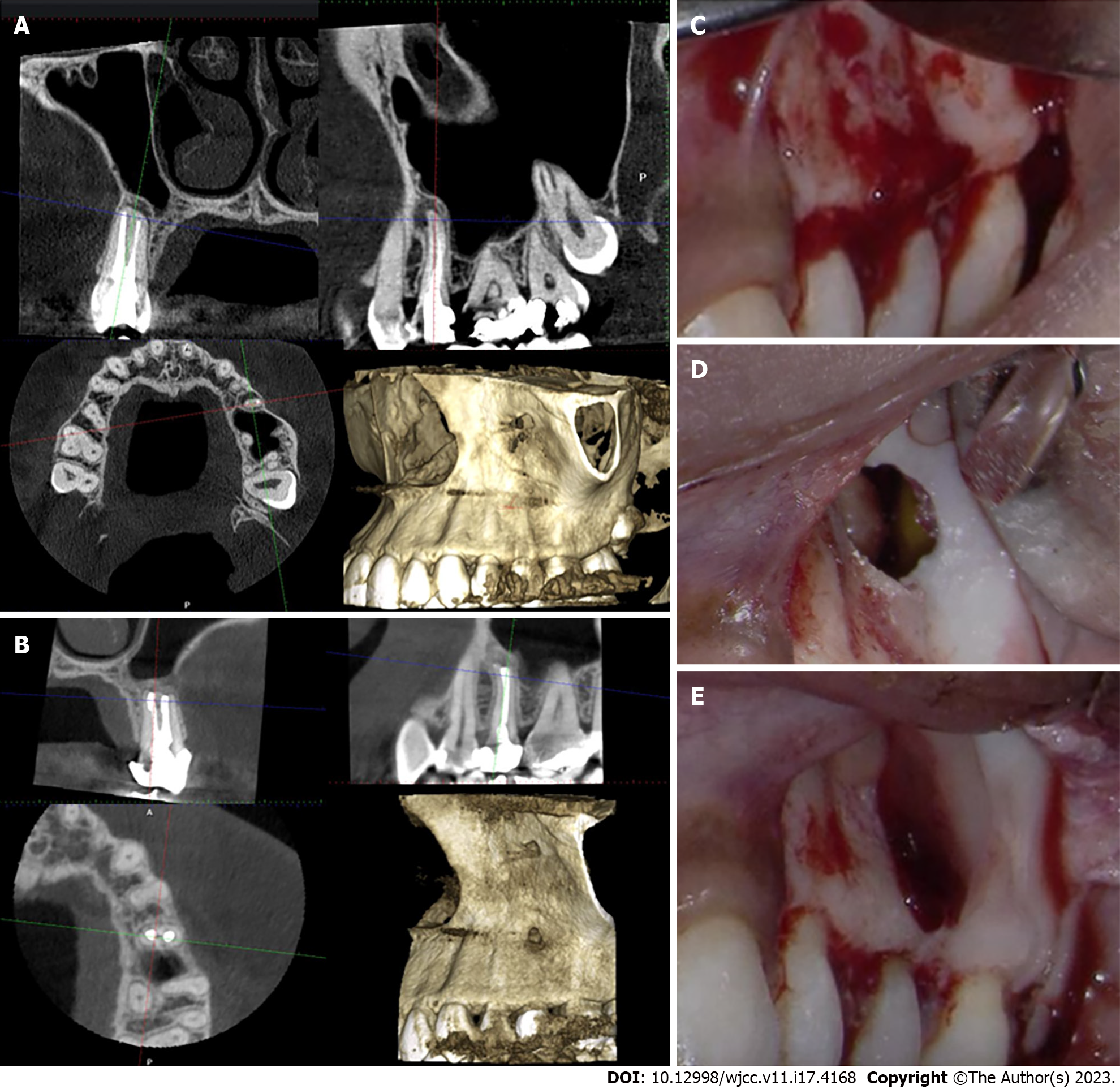
Figure 2 Periapical endodontic surgery on the maxillary left second premolar using advanced platelet-rich fibrin membrane.
A: The cone-beam computed tomography (CBCT) shows localized radiolucency surrounding the apex of the maxillary left second premolar in the sagittal section. There is a thin layer of the labial bone surrounding the apex and a displaced sinus periosteum upward which is a typical feature of periapical osteoperiostitis in the sagittal section. The root canal treatment appears to be adequate with a uniform density; B: The CBCT shows bony healing of the resected area within four months and a normal appearance of the maxillary sinus; C: A clinical photo of the labial bone before root resection; D: A clinical photo of the bony defect after resection shows a hollow space adjacent to the maxillary sinus; E: The platelet-rich fibrin membrane is used to seal the bony defect.
Figure 3 Periapical endodontic surgery on the maxillary right lateral incisor advanced platelet-rich fibrin membrane.
A: The cone-beam computed tomography (CBCT) shows a well-defined radiolucency surrounding the apex of the maxillary right lateral anterior in the sagittal section. The lesion appears to be perforating the labial and palatal bone surrounding the apex in the coronal section. The root canal appears to be adequate and uniform in density; B: The CBCT shows the bone formation and healing surrounding the apex and bony defect within four months post-operatively in the coronal and sagittal section; C: A clinical photo shows the resected surface of the root and the bony defect; D: The placement of bone graft mixed with the blood clot; E: The platelet-rich fibrin membrane is used to seal the defect and hold the bone graft in place.
- Citation: Algahtani FN, Almohareb R, Aljamie M, Alkhunaini N, ALHarthi SS, Barakat R. Application of advanced platelet-rich fibrin for through-and-through bony defect during endodontic surgery: Three case reports and review of the literature. World J Clin Cases 2023; 11(17): 4168-4178
- URL: https://www.wjgnet.com/2307-8960/full/v11/i17/4168.htm
- DOI: https://dx.doi.org/10.12998/wjcc.v11.i17.4168


