Copyright
©The Author(s) 2023.
World J Clin Cases. May 16, 2023; 11(14): 3311-3316
Published online May 16, 2023. doi: 10.12998/wjcc.v11.i14.3311
Published online May 16, 2023. doi: 10.12998/wjcc.v11.i14.3311
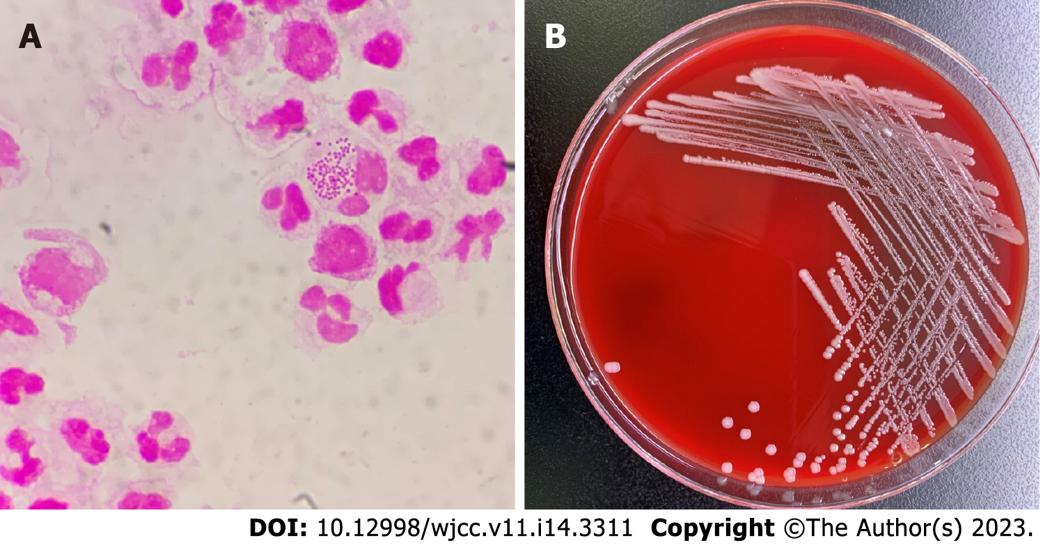
Figure 1 Smear examination and bacterial culture results of peritoneal dialysis fluid.
A: Gram staining of peritoneal dialysate specimens after centrifugation (×1000); B: Colony morphology on a blood agar plate at 35 °C, 5% CO2 and cultured for 48 h.
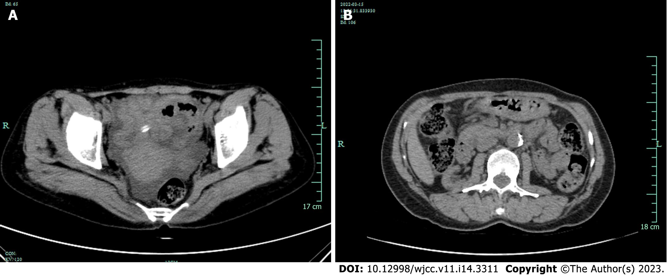
Figure 2 Computed tomography results before and after treatment.
A: Before treatment, computed tomography (CT) showed ascites without obvious thickening of the membrane; B: CT reexamination showed that peritoneal thickening has not been completely absorbed caused by peritonitis after treatment.
- Citation: Ren JM, Zhang XY, Liu SY. Neisseria mucosa - A rare cause of peritoneal dialysis-related peritonitis: A case report. World J Clin Cases 2023; 11(14): 3311-3316
- URL: https://www.wjgnet.com/2307-8960/full/v11/i14/3311.htm
- DOI: https://dx.doi.org/10.12998/wjcc.v11.i14.3311


