Copyright
©The Author(s) 2023.
World J Clin Cases. May 16, 2023; 11(14): 3295-3303
Published online May 16, 2023. doi: 10.12998/wjcc.v11.i14.3295
Published online May 16, 2023. doi: 10.12998/wjcc.v11.i14.3295
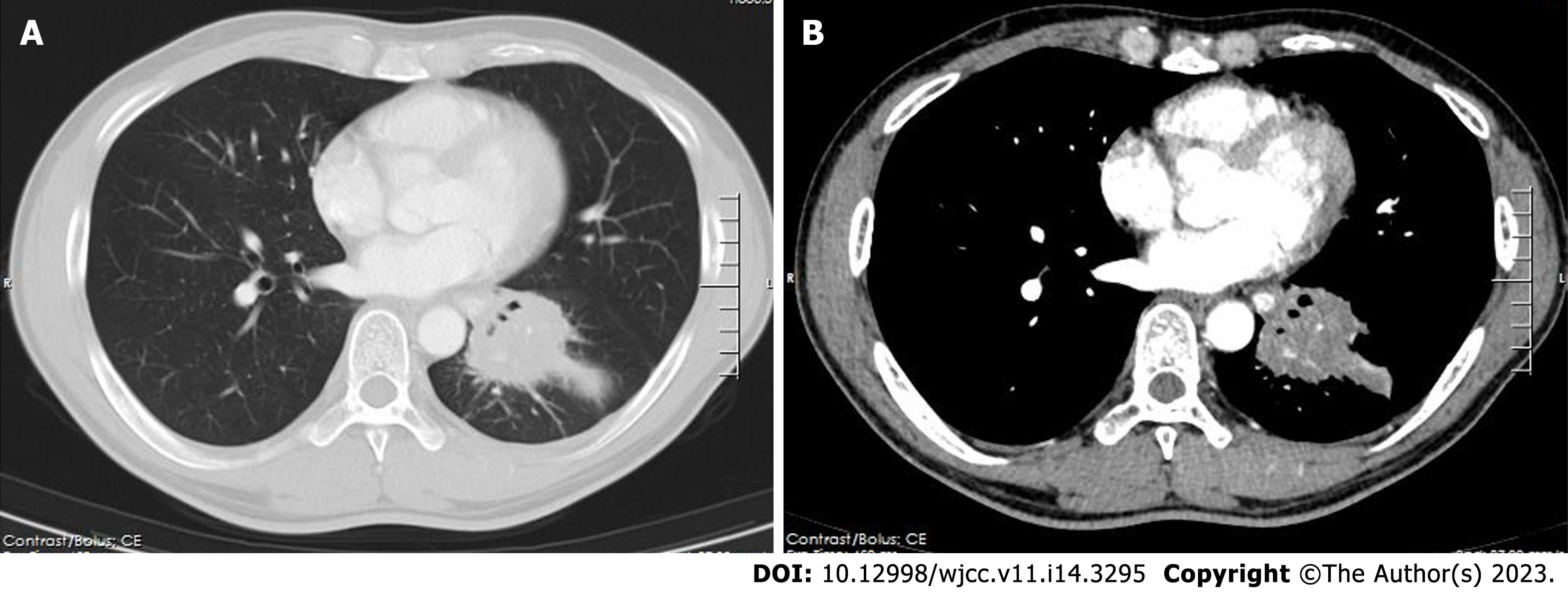
Figure 1 High-resolution chest contrast-enhanced computed tomography.
A: Lung window; and B: Mediastinal window. A mass in the left lower lobe with a diameter of approximately 6 cm. It was slightly enhanced and mainly located in the lateral segment (S9) and posterior segment (S10). The pulmonary vasculature was still looming in this mass, but there was bronchial occlusion of the affected lung segments.
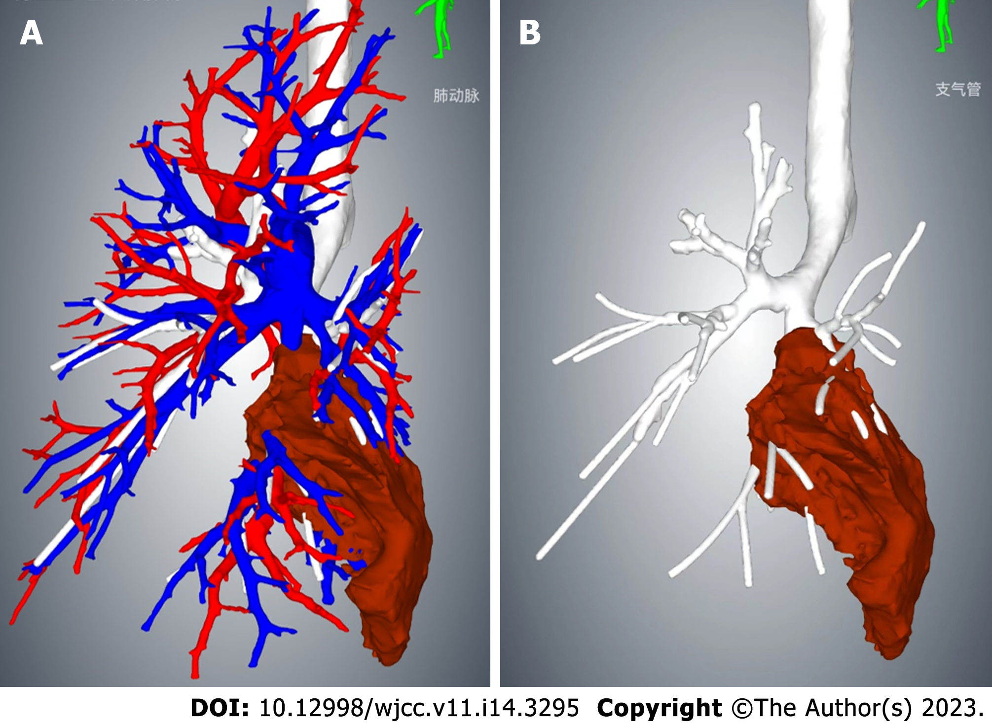
Figure 2 Three-dimensional reconstruction of the chest based on computed tomography images.
A: The pulmonary vasculature is visible; and B: The pulmonary vasculature is hidden. The mass volume calculated by 3D reconstruction was 136 mL.
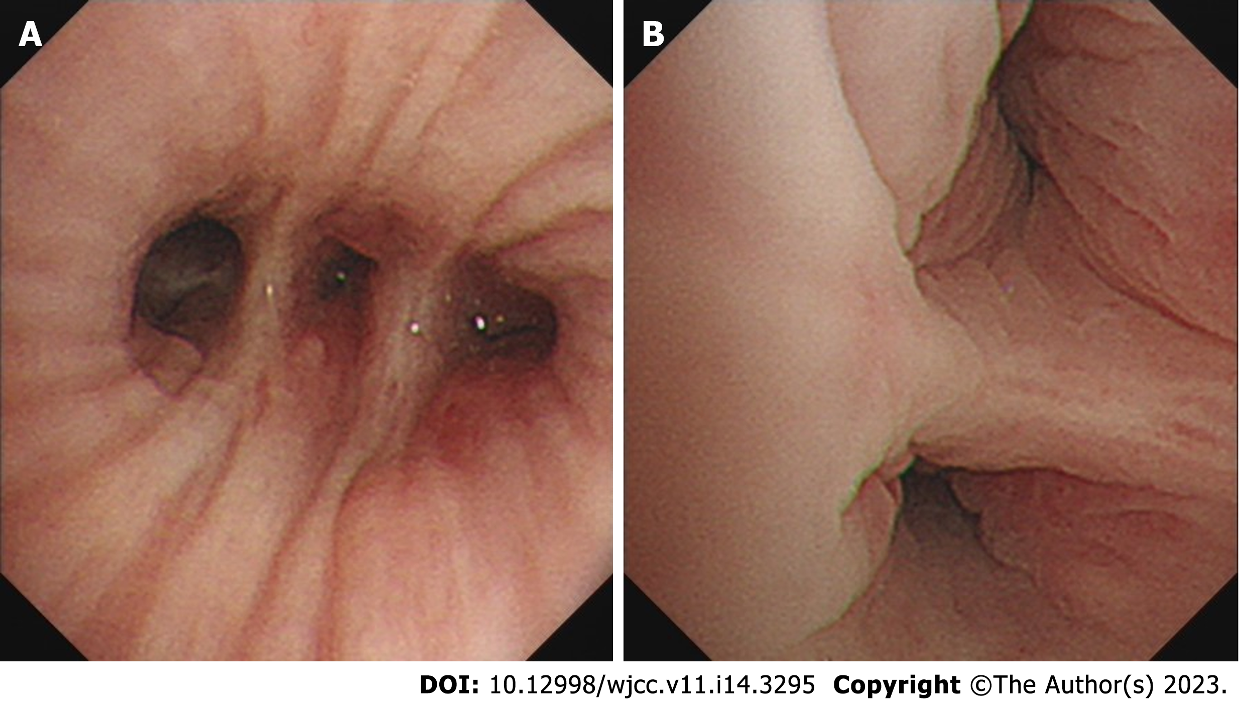
Figure 3 Images during electronic bronchoscopy.
A: Bronchoscopy revealed that the lumen of the left lower lobe's lateral and posterior segmental bronchi was narrowed; and B: The mucosa was congested and swollen, and no new organisms were found in the lumen.
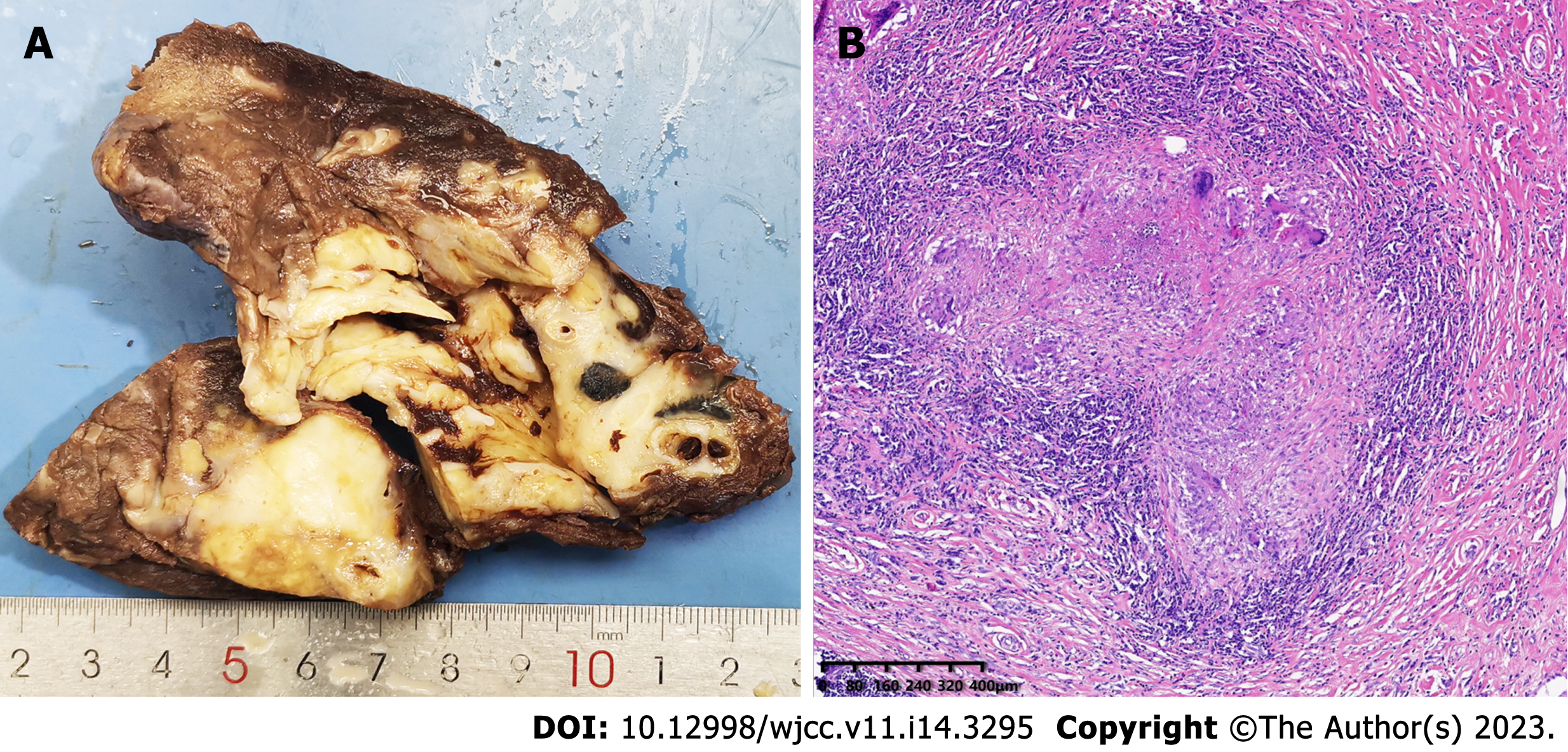
Figure 4 Macroscopic and microscopic features of the left lower lobe mass.
A: The mass was firm, approximately 6 cm in diameter, and pale in color when sectioned; and B: Hematoxylin and eosin stained section revealed that the left lower lobe mass was an inflammatory granuloma. Interstitial fibrous tissue hyperplasia, inflammatory cell infiltration, and multinucleated giant cell reaction were all observed under the microscope.
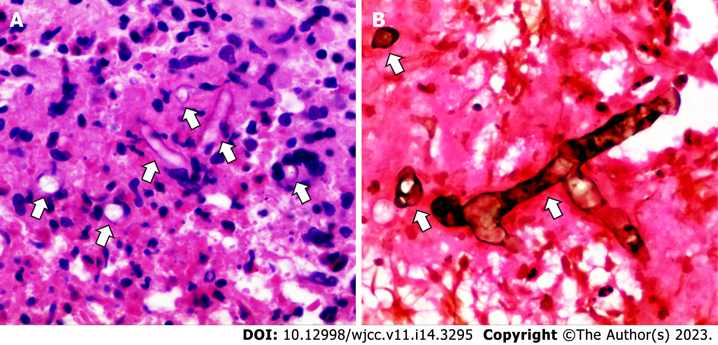
Figure 5 Postoperative histopathological results of the left lower lobe mass.
A: Hematoxylin and eosin staining (400 ×); and B: Hexamine silver staining (400 ×), showed broad, right-angled branched aseptate hyphae indicating mucormycosis (marked with white arrows).
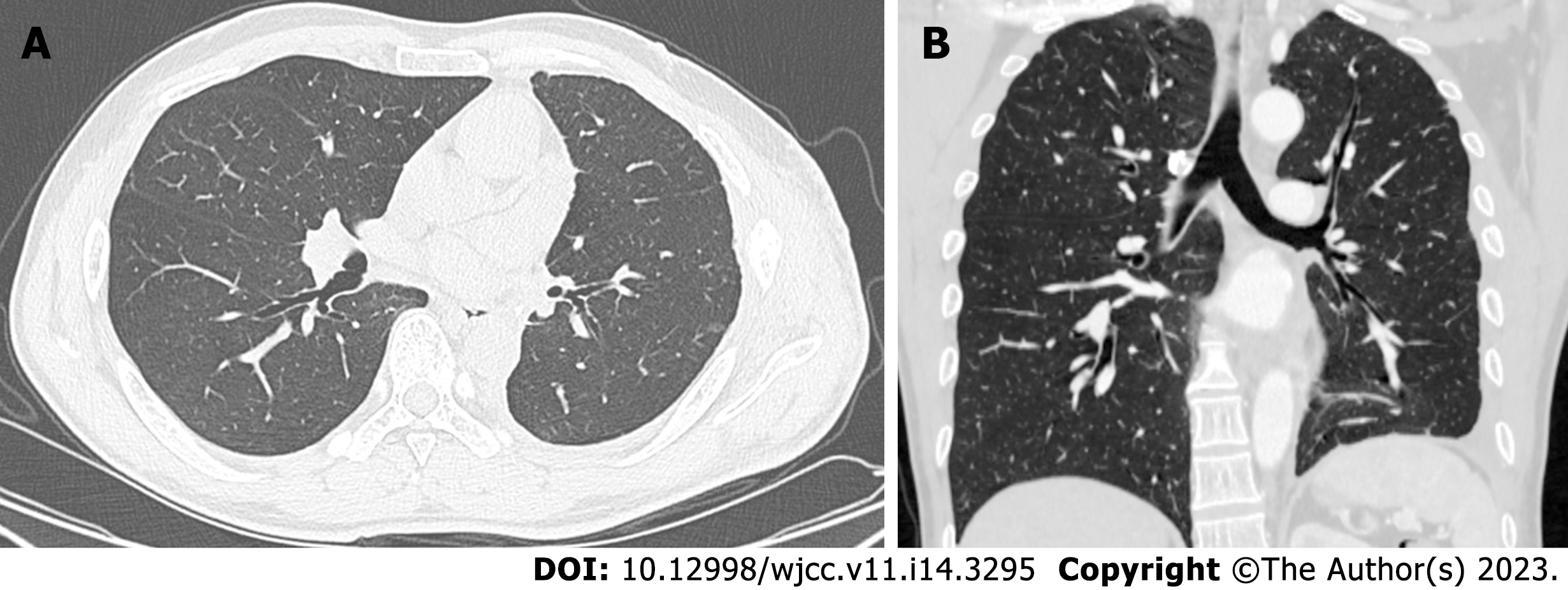
Figure 6 High-resolution chest contrast-enhanced computed tomography.
A: Transverse plane; and B: Coronal plane. A re-examination of computed tomography three months after the operation showed that the lungs were in good condition, and no recurrence was found.
- Citation: Guo XZ, Gong LH, Wang WX, Yang DS, Zhang BH, Zhou ZT, Yu XH. Chronic pulmonary mucormycosis caused by rhizopus microsporus mimics lung carcinoma in an immunocompetent adult: A case report. World J Clin Cases 2023; 11(14): 3295-3303
- URL: https://www.wjgnet.com/2307-8960/full/v11/i14/3295.htm
- DOI: https://dx.doi.org/10.12998/wjcc.v11.i14.3295


