Copyright
©The Author(s) 2023.
World J Clin Cases. May 16, 2023; 11(14): 3267-3274
Published online May 16, 2023. doi: 10.12998/wjcc.v11.i14.3267
Published online May 16, 2023. doi: 10.12998/wjcc.v11.i14.3267
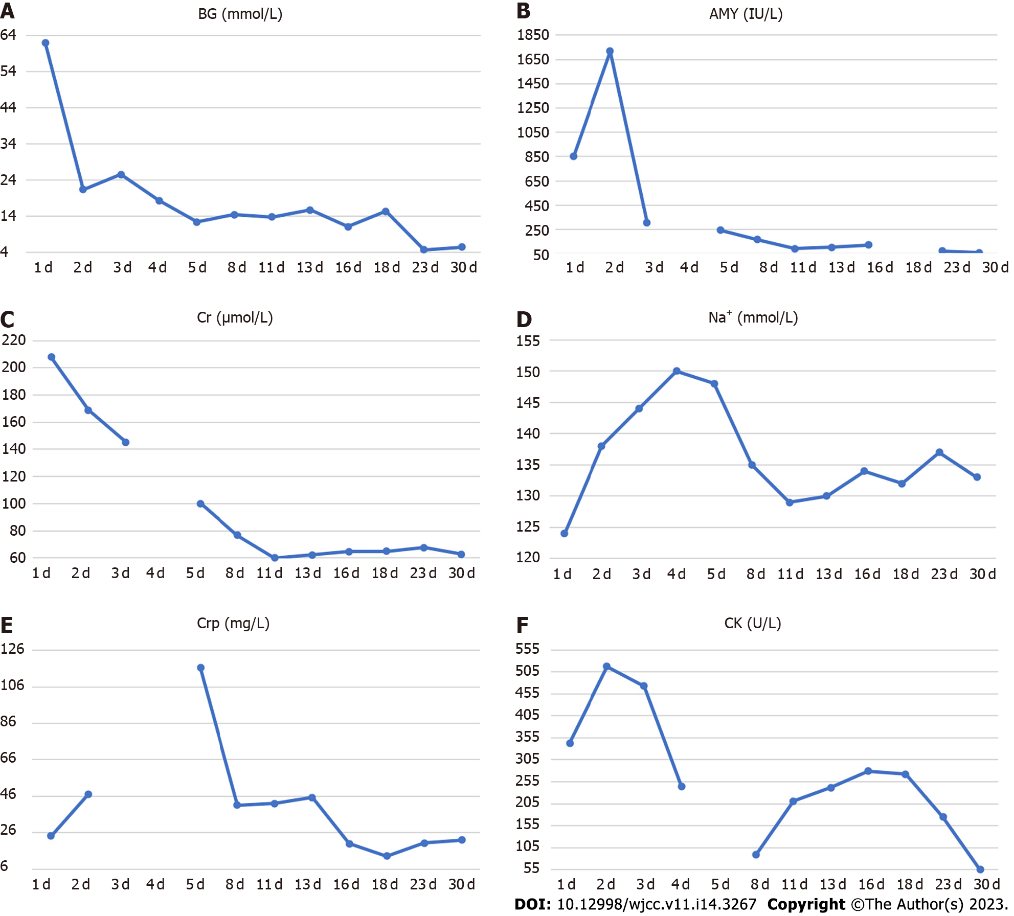
Figure 1 Temporal changes in laboratory parameters during treatment and follow-up.
A: Blood glucose; B: Amylase; C: Serum creatinine; D: Serum sodium; E: C-reactive protein; F: Creatine kinase.
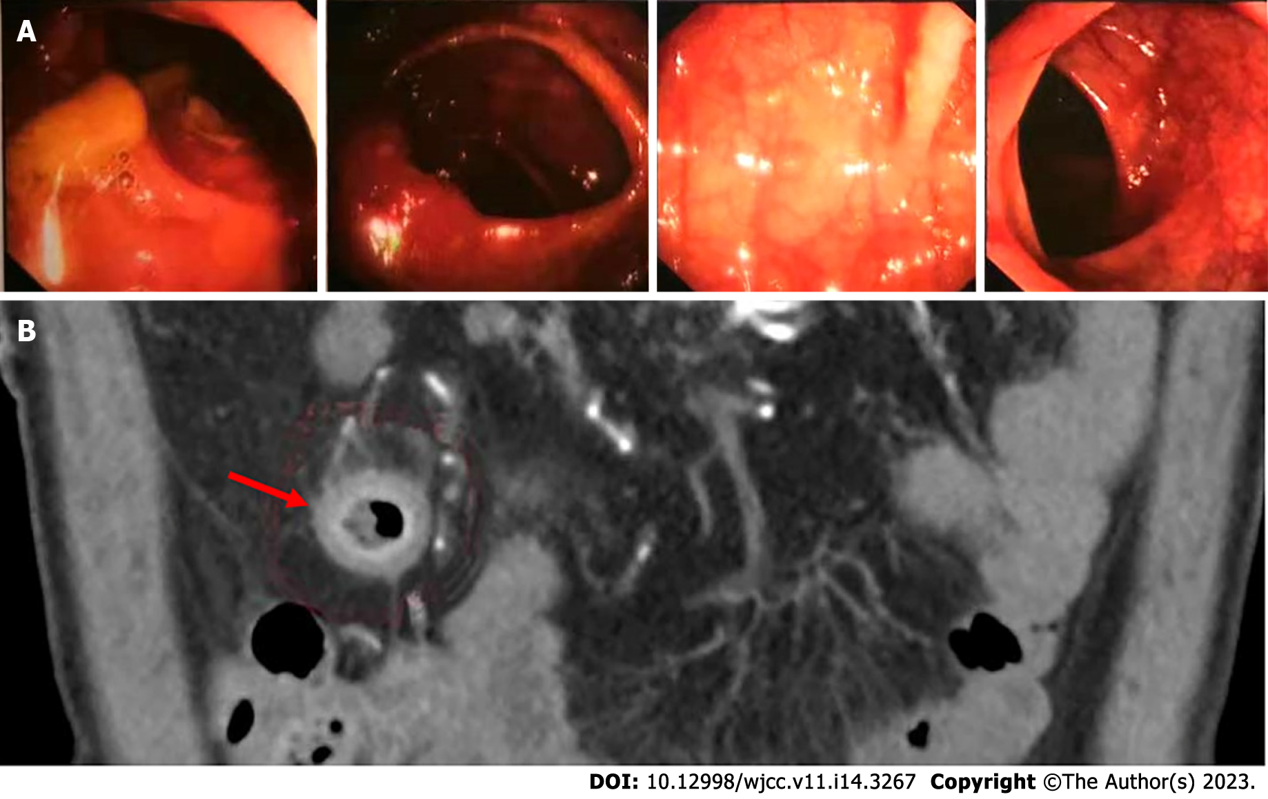
Figure 2 Colonoscopy and abdominal computed tomography examination.
A: Electronic colonoscopy image; B: Abdominal computed tomography scan.
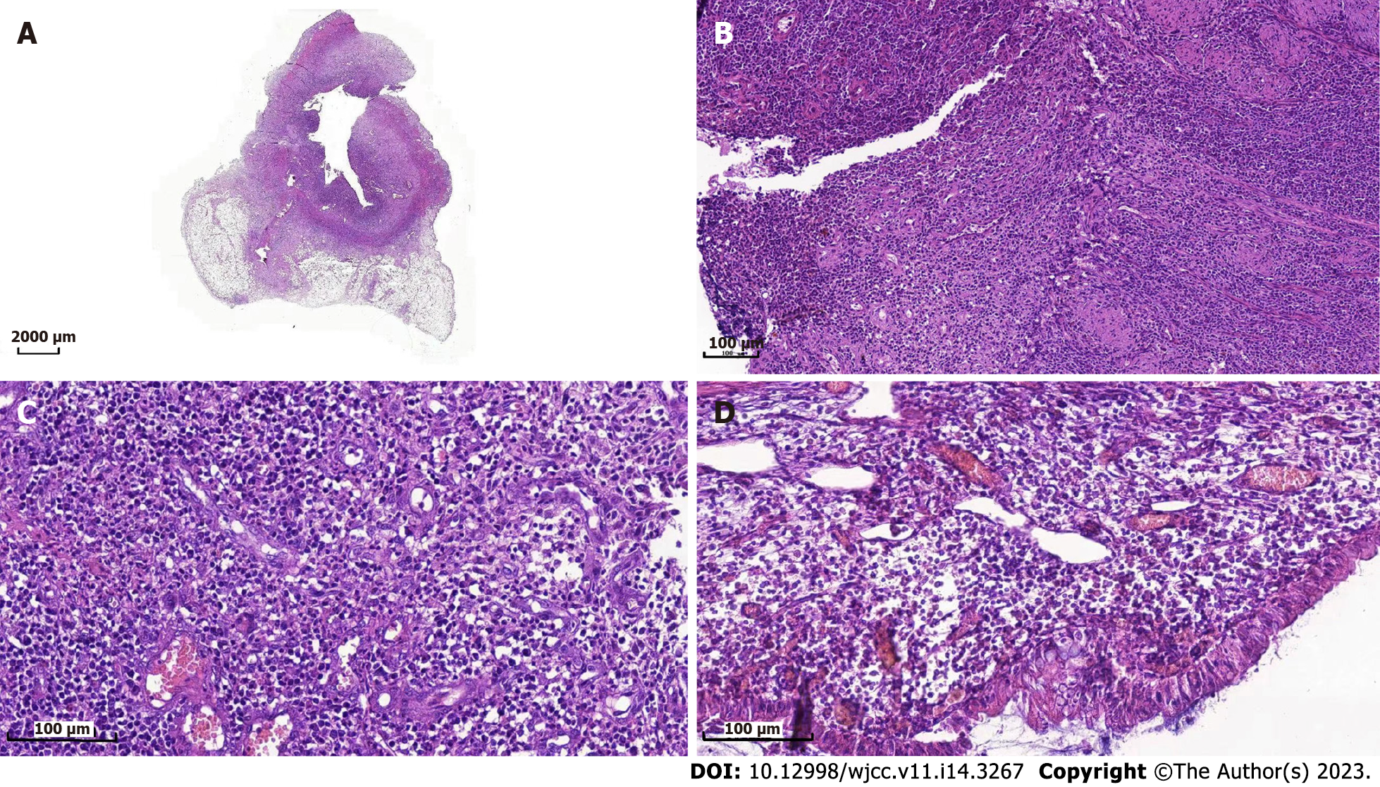
Figure 3 Hematoxylin and eosin-stained sections of terminal ileum.
A: 5 times amplification by microscope of the lesions in intestine; B: 50 times amplification by microscope of the lesions in intestine; C: 100 times amplification by microscope of the lesions in intestine; D: 200 times amplification by microscope of the lesions in intestine.
- Citation: Gao MJ, Xu Y, Wang WB. Immune checkpoint inhibitor therapy-induced autoimmune polyendocrine syndrome type II and Crohn’s disease: A case report. World J Clin Cases 2023; 11(14): 3267-3274
- URL: https://www.wjgnet.com/2307-8960/full/v11/i14/3267.htm
- DOI: https://dx.doi.org/10.12998/wjcc.v11.i14.3267


