Copyright
©The Author(s) 2023.
World J Clin Cases. May 16, 2023; 11(14): 3167-3175
Published online May 16, 2023. doi: 10.12998/wjcc.v11.i14.3167
Published online May 16, 2023. doi: 10.12998/wjcc.v11.i14.3167
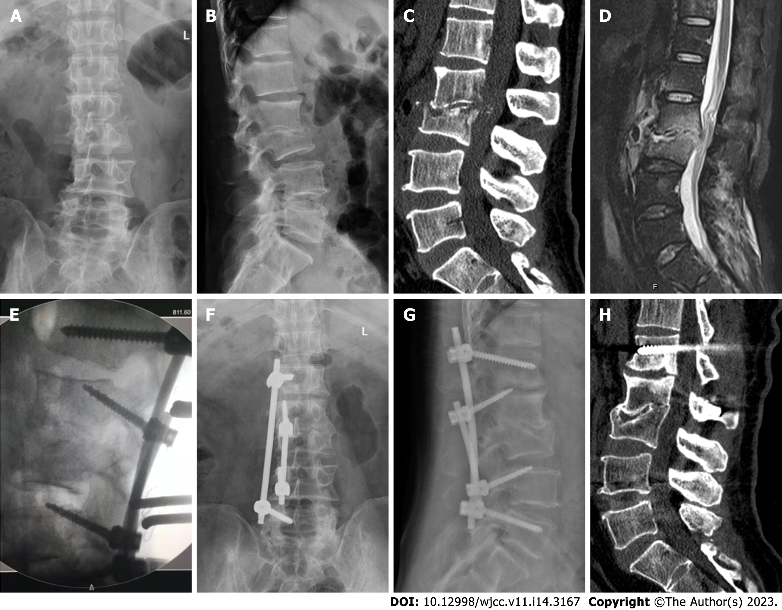
Figure 1 A typical case with bone fusion.
A 47-year-old male patient presented with low back pain and left leg numbness and weakness. A and B: The pathological diagnosis was L2/3 lumbar tuberculosis, preoperative lumbar spinal X-ray showed L2/3 intervertebral space collapse; C: Lumbar spinal CT showed L2/3 intervertebral space stenosis and vertebral body destruction, and sequestrum could be seen invading the front of the intervertebral space and protruding backwards into the spinal canal; D: Fat-suppressed magnetic resonance imaging of the lumbar vertebrae showed destruction of the L2/3 intervertebral disc, narrowing of the intervertebral space, pus formation in the intervertebral space protruding towards the front and back of the intervertebral space, and compression of the dural sac; E: C-arm fluoroscopy during the operation confirmed good positioning of the pedicle screws and cortical bone trajectory screws; F and G: X-ray of the lumbar spine 1 year after the operation showed L2/3 intervertebral space fusion, normal positioning of the internal fixation device, and the absence of broken screws and rods; and H: CT scan of the lumbar spine 1 year after the operation showed that the L2/3 vertebral bodies had fused and that tuberculosis lesions had not developed. CT: Computed tomography.
- Citation: Yuan YF, Ren ZX, Zhang C, Li GJ, Liu BZ, Li XD, Miao J, Li JF. Multitrack and multianchor point screw technique combined with the Wiltse approach for lesion debridement for lumbar tuberculosis. World J Clin Cases 2023; 11(14): 3167-3175
- URL: https://www.wjgnet.com/2307-8960/full/v11/i14/3167.htm
- DOI: https://dx.doi.org/10.12998/wjcc.v11.i14.3167


