Copyright
©The Author(s) 2023.
World J Clin Cases. May 6, 2023; 11(13): 3045-3051
Published online May 6, 2023. doi: 10.12998/wjcc.v11.i13.3045
Published online May 6, 2023. doi: 10.12998/wjcc.v11.i13.3045
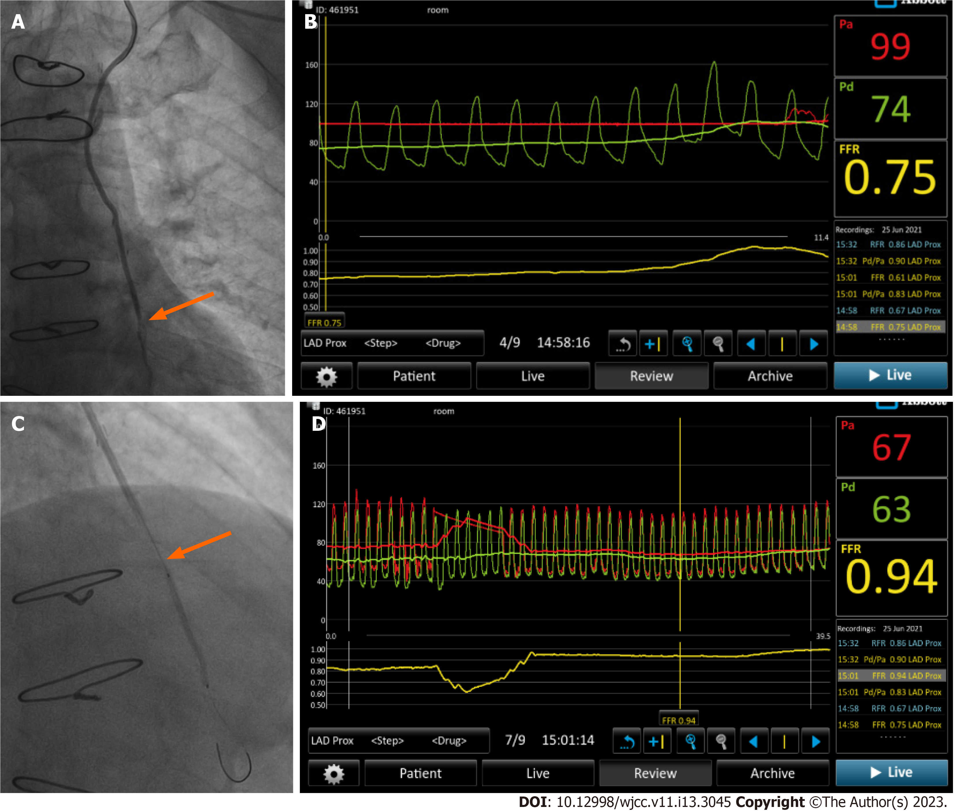
Figure 1 Images of case 1.
A: Angiography showed 85% stenosis at the distal end of the anastomosis of the left internal mammary artery (LIMA)-left anterior descending artery graft (orange arrow); B: Before intervention, the fractional flow reserve measured via the LIMA was 0.75 (positive); C: Dilation with a 2.0 mm × 31.0 mm drug containing balloon was performed at the stenotic segment of the left anterior descending artery via the LIMA (orange arrow); D: After balloon dilation, remeasurement of pressure showed that the fractional flow reserve was 0.94.
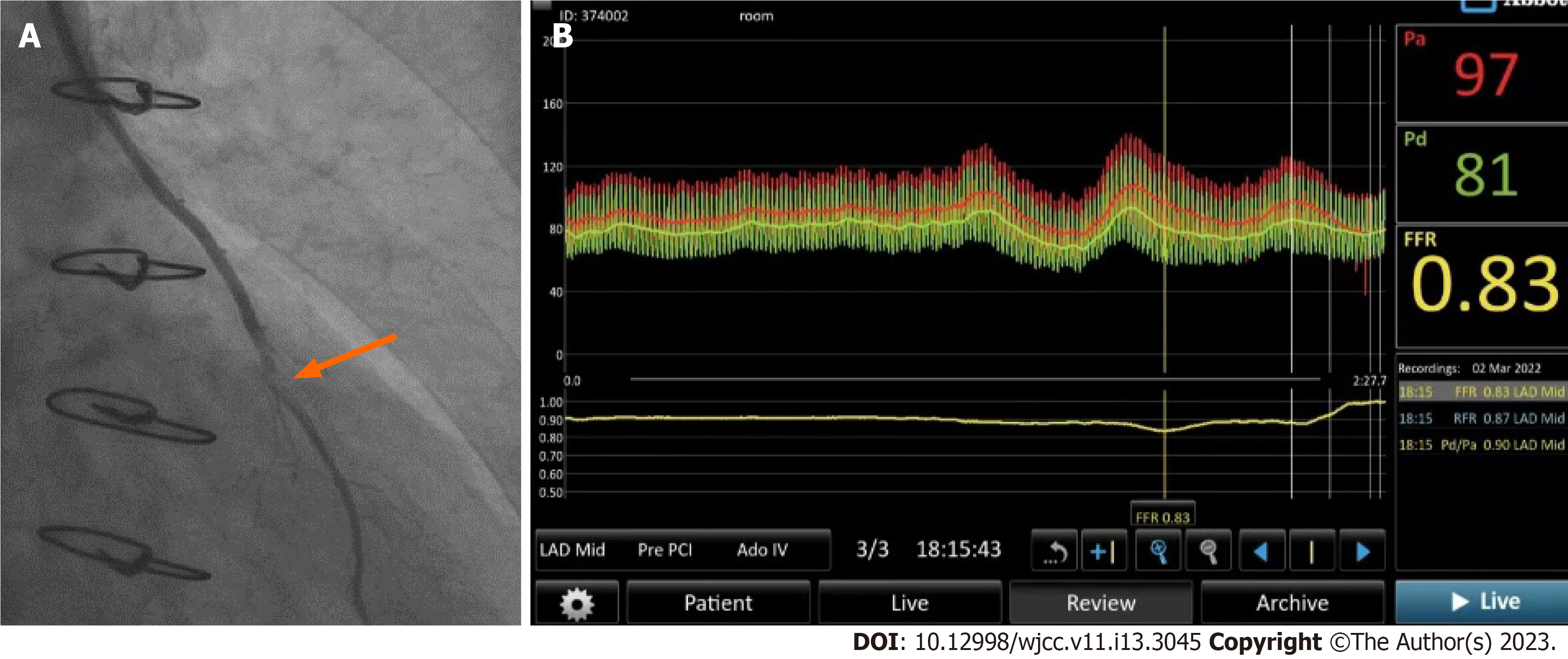
Figure 2 Images of case 2.
A: Angiography indicated 75% anastomotic stenosis (orange arrow); B: The fractional flow reserve measured from the left internal mammary artery to the distal end of the anastomosis was 0.83 (negative).
- Citation: Zhang LY, Gan YR, Wang YZ, Xie DX, Kou ZK, Kou XQ, Zhang YL, Li B, Mao R, Liang TX, Xie J, Jin JJ, Yang JM. Fractional flow reserve measured via left internal mammary artery after coronary artery bypass grafting: Two case reports. World J Clin Cases 2023; 11(13): 3045-3051
- URL: https://www.wjgnet.com/2307-8960/full/v11/i13/3045.htm
- DOI: https://dx.doi.org/10.12998/wjcc.v11.i13.3045


