Copyright
©The Author(s) 2023.
World J Clin Cases. May 6, 2023; 11(13): 3022-3028
Published online May 6, 2023. doi: 10.12998/wjcc.v11.i13.3022
Published online May 6, 2023. doi: 10.12998/wjcc.v11.i13.3022
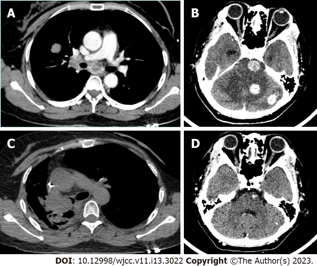
Figure 1 Computed tomography findings of the patient's lungs and brain.
A: Computed tomography (CT) examination in May 2021 showed that there was a space-occupying lesion in the lung lobe, and no obvious mass was seen in the right hilum; B: CT examination in May 2021 showed three intracranial space-occupying lesions; C: CT examination in November 2021 showed a soft tissue mass in the right hilum, blocking the bronchial lumen; D: CT examination in November 2021 indicated that the nodules in the brain had shrunk or disappeared.
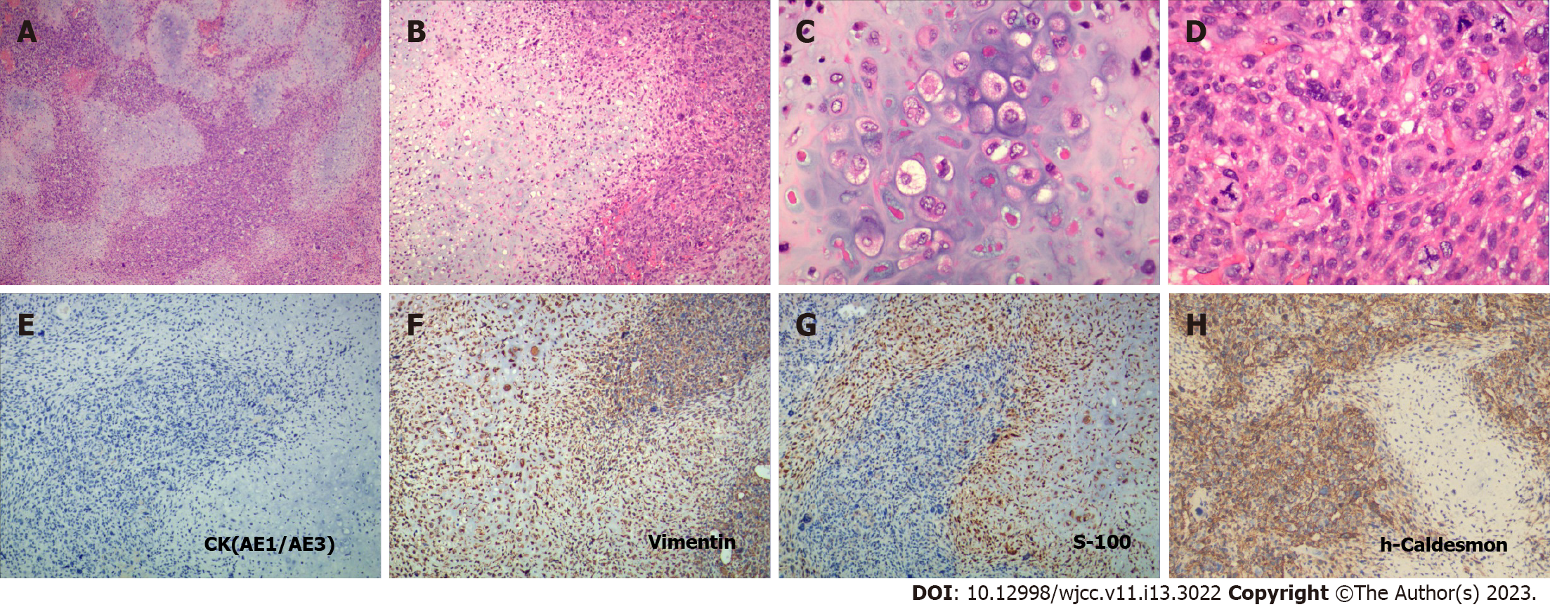
Figure 2 Histopathologic and immunohistochemical observation of the right hilum mass.
A and B: Dedifferentiated chondrosarcoma under low magnification and medium magnification, respectively, showed the tumors characterized by the following two distinct histopathologic components: a low-grade chondrosarcoma region sharply juxtaposed with a high-grade noncartilaginous sarcoma component; C: Typical low-grade chondrosarcoma region; D: Typical high-grade dedifferentiated sarcoma region; E: Negative expression of epithelial marker Cytokeratin in dedifferentiated chondrosarcoma (DDCS); F: Positive expression of mesenchymal marker Vimentin in DDCS; G: Positive expression of S-100 in low-grade chondrosarcoma region; H: Expression of h-Caldesmon in high-grade dedifferentiated sarcoma region, suggesting that the dedifferentiated sarcoma may have smooth muscle cell differentiation (A to D: Hematoxylin and eosin staining, A: × 40, B: × 100, C and D: × 400; E to H: Immunohistochemical staining, × 100).
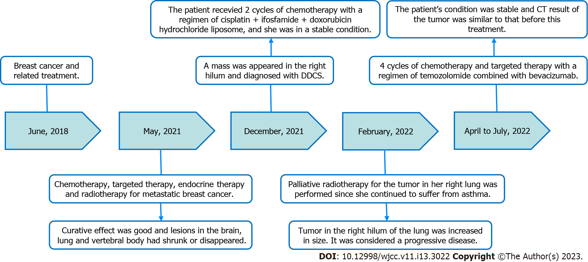
Figure 3 Timeline summarizing the main treatment and outcome of this case report.
CT: Computed tomography; DDCS: Dedifferentiated chondrosarcoma.
- Citation: Wen H, Gong FJ, Xi JM. Primary dedifferentiated chondrosarcoma of the lung with a 4-year history of breast cancer: A case report. World J Clin Cases 2023; 11(13): 3022-3028
- URL: https://www.wjgnet.com/2307-8960/full/v11/i13/3022.htm
- DOI: https://dx.doi.org/10.12998/wjcc.v11.i13.3022


