Copyright
©The Author(s) 2023.
World J Clin Cases. Apr 26, 2023; 11(12): 2694-2707
Published online Apr 26, 2023. doi: 10.12998/wjcc.v11.i12.2694
Published online Apr 26, 2023. doi: 10.12998/wjcc.v11.i12.2694
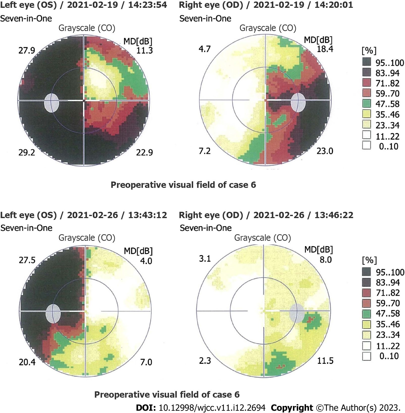
Figure 1 Comparison of the preoperative and postoperative visual fields for Case 6.
Preoperative visual field examination showed bitemporal hemianopsia, which was more severe in the left eye. Three days after surgery, examination revealed a partial temporal visual field defect in the left eye and a standard visual field in the right eye.
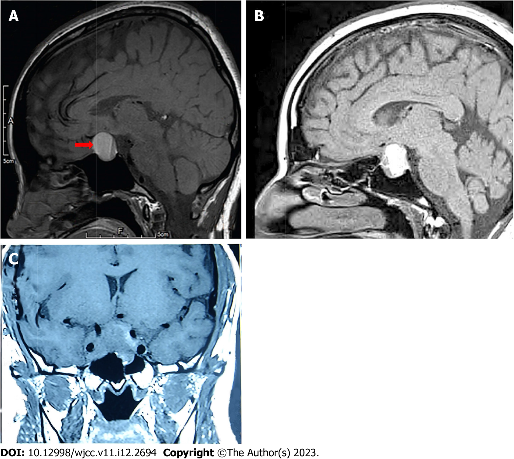
Figure 2 Three typical images of pituitary adenoma apoplexy.
A: Sagittal T1 weighted imaging (T1WI) showed isointensity and hyperintensity with a visible liquid level (Case 15); B: Sagittal T1WI showed mixed intensity (mainly hyperintensity) (Case 40); C: Coronal T1WI showed isointensity (Case 5).
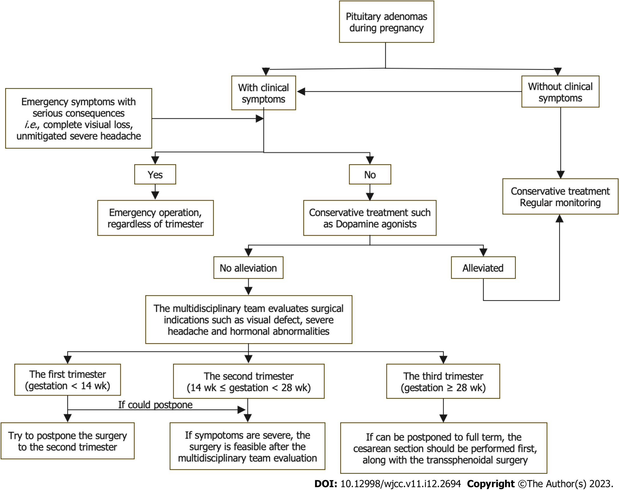
Figure 3 Flow diagram of treatment procedures for pituitary adenoma during pregnancy.
- Citation: Jia XY, Guo XP, Yao Y, Deng K, Lian W, Xing B. Surgical management of pituitary adenoma during pregnancy. World J Clin Cases 2023; 11(12): 2694-2707
- URL: https://www.wjgnet.com/2307-8960/full/v11/i12/2694.htm
- DOI: https://dx.doi.org/10.12998/wjcc.v11.i12.2694


