Copyright
©The Author(s) 2023.
World J Clin Cases. Apr 16, 2023; 11(11): 2576-2581
Published online Apr 16, 2023. doi: 10.12998/wjcc.v11.i11.2576
Published online Apr 16, 2023. doi: 10.12998/wjcc.v11.i11.2576
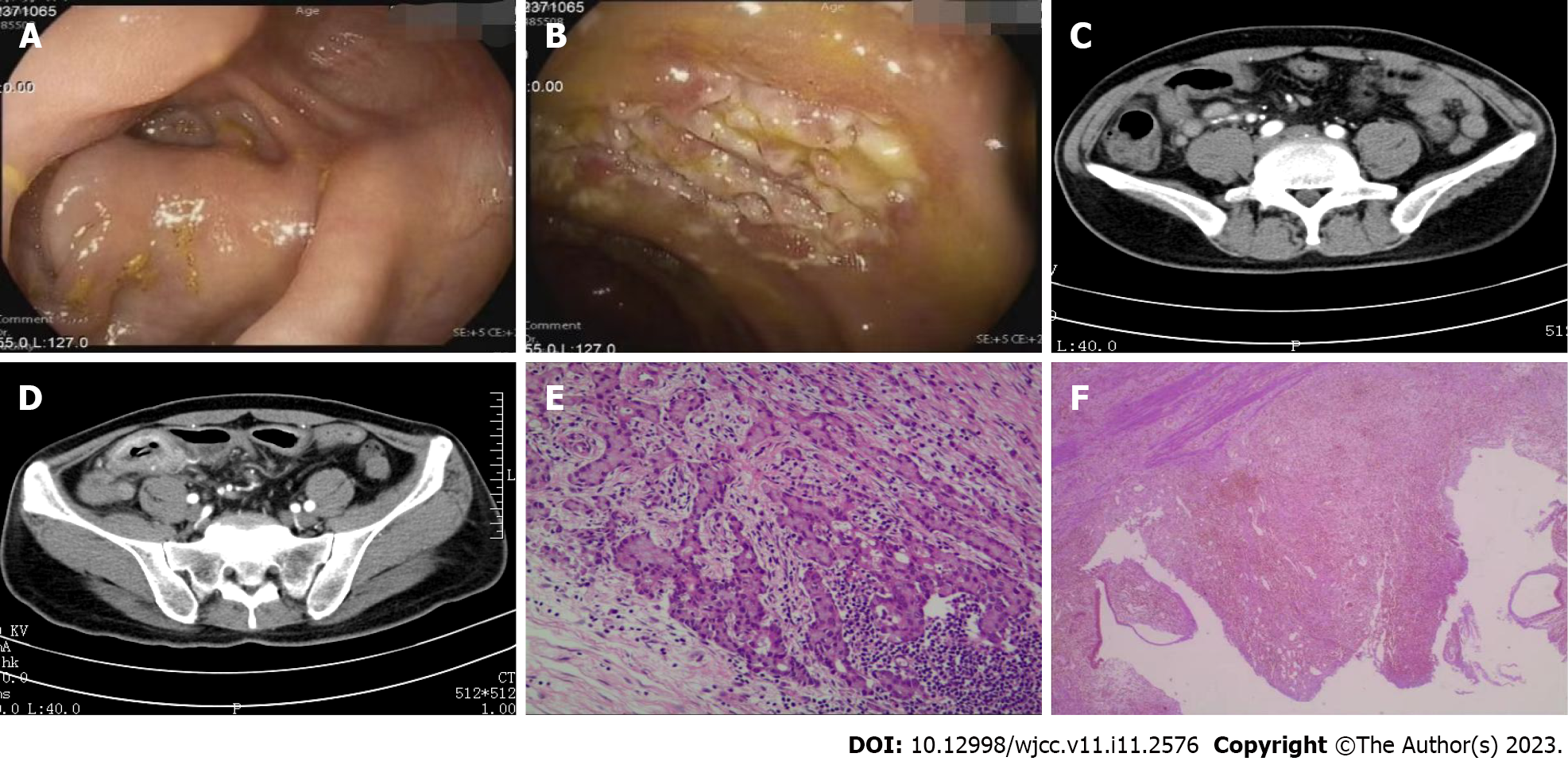
Figure 1 Preoperative colonoscopy, computed tomography and postoperative pathological images.
A: Ileocecum with poor exposure of the appendiceal foramen; B: Multiple ulcers in the terminal ileum; C: Multiple enlarged lymph nodes in the abdomen; D: The wall of the distal ileum was thickened with abnormal enhancement, and multiple adjacent, enlarged lymph nodes; E: Postoperative pathologic evaluation indicated high-grade goblet cell adenocarcinoma of the appendix (original magnification × 400); F: The terminal ileum indicated chronic active inflammation (original magnification × 400).
- Citation: Mao YH, Li L, Wen LM, Qin JM, Yang YL, Wang L, Wang FR, Zhao YZ. Autoimmune encephalitis after surgery for appendiceal cancer: A case report. World J Clin Cases 2023; 11(11): 2576-2581
- URL: https://www.wjgnet.com/2307-8960/full/v11/i11/2576.htm
- DOI: https://dx.doi.org/10.12998/wjcc.v11.i11.2576


