Copyright
©The Author(s) 2023.
World J Clin Cases. Apr 16, 2023; 11(11): 2559-2566
Published online Apr 16, 2023. doi: 10.12998/wjcc.v11.i11.2559
Published online Apr 16, 2023. doi: 10.12998/wjcc.v11.i11.2559
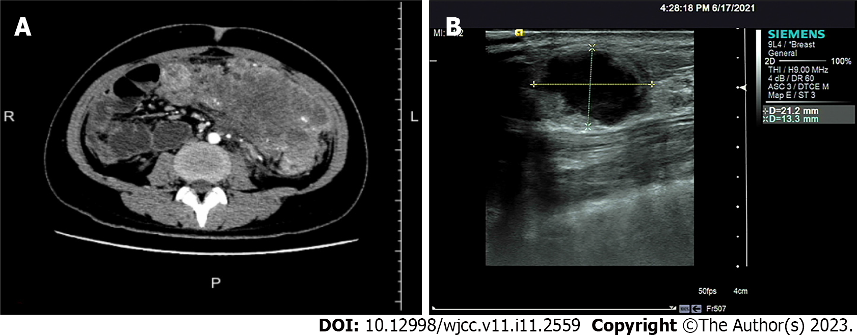
Figure 1 Imaging findings of the abdominal tumor and breast tumor.
A: Abdominal computed tomography showed that the abdominal tumor measured 7.5 cm × 10.9 cm, with uneven density, and the boundary between the tumor and the surrounding tissues was unclear; B: Breast ultrasound clearly showed the breast tumor measuring 2.1 cm × 1.5 cm × 1.3 cm, with an unclear boundary and echo heterogenicity. Color Doppler flow imaging showed short rod blood flow signal.
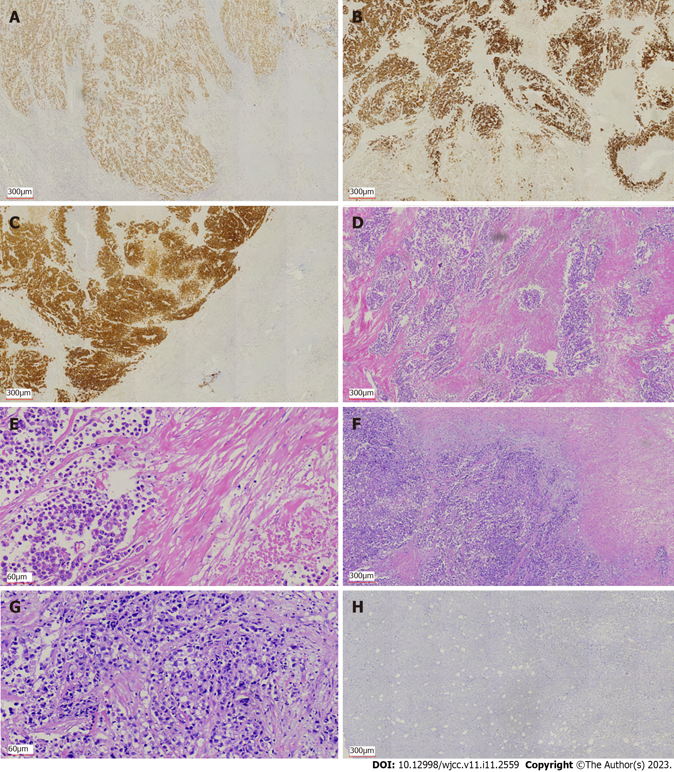
Figure 2 Immunohistochemical and histological features of the primary colon cancer and breast metastasis from colon cancer.
A-C: Immunohistochemical staining showed expression of caudal-related homeobox transcription factor 2 (A), mucin-2 (B), and villin (C) in colon cancer; D and E: Hematoxylin and eosin (H&E) staining showed intracellular mucus in colon cancer (D, 40 ×; E, 200 ×); F: H&E staining showed breast metastasis from colon cancer (40 ×); G: H&E staining showed adenocarcinoma cells in the breast metastasis (200 ×); H: Immunohistochemical staining showed negative expression of GATA binding protein-3 in breast metastasis from colon cancer.
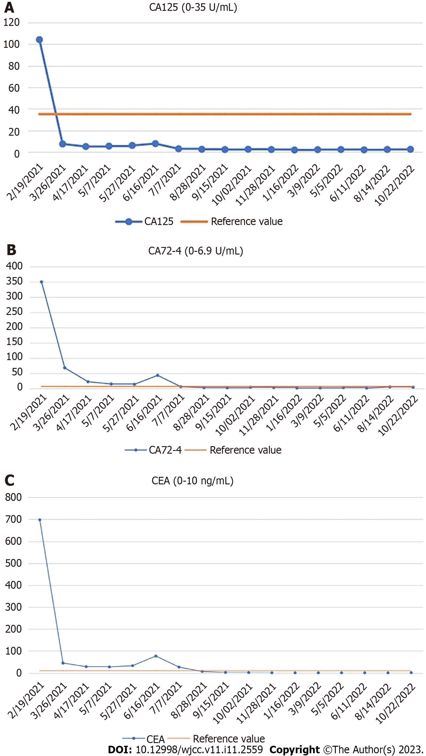
Figure 3 Changes in serum levels of carcinoembryonic antigen, carbohydrate antigen 72-4, and carbohydrate antigen 125.
A: Carbohydrate antigen (CA) 125 (0-35 U/mL); B: CA72-4 (0-6.9 U/mL); C: Carcinoembryonic antigen (0-10 ng/mL). CA: Carbohydrate antigen; CEA: Carcinoembryonic antigen.
- Citation: Jiao X, Xing FZ, Zhai MM, Sun P. Successful treatment of breast metastasis from primary transverse colon cancer: A case report. World J Clin Cases 2023; 11(11): 2559-2566
- URL: https://www.wjgnet.com/2307-8960/full/v11/i11/2559.htm
- DOI: https://dx.doi.org/10.12998/wjcc.v11.i11.2559


